
Page 904 - Hematology_ Basic Principles and Practice ( PDFDrive )
P. 904
Chapter 56 Conventional and Molecular Cytogenomic Basis of Hematologic Malignancies 787
CHR 9
DER(22)
CHR 22
22
9 DER(9)
A B
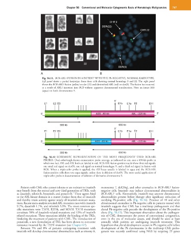
Fig. 56.11. BCR-ABL1 FUSION IN A PATIENT WITH THE Ph-NEGATIVE, NORMAL KARYOTYPE.
Left panel shows a partial karyotype from three cells showing normal homologs 9 and 22. The right panel
shows the BCR-ABL1 fusion (yellow) on der (22) and diminished ABL (red) on der(9). The fusion has occurred
as a result of ABL1 insertion into BCR without apparent chromosomal translocation. Note an intact ASS
(aqua) on both chromosomes 9.
5'
E1 E2 E3 E1b E1a E2 E11
5' ASS ~65 kb ABL ~225 kb 3'
300 kb 650 kb
5'
E1 E2 E3 E1b E1a E2 E11
5' ASS ~65 kb ABL ~225 kb 3'
300 kb 650 kb
Fig. 56.12 SCHEMATIC REPRESENTATION OF THE MOST FREQUENTLY USED BCR-ABL
PROBES. Dual-color/single-fusion extrasensitive probe strategy, as indicated in text, uses a 650-kb probe in
which two loci, ABL and ASS, both are labeled in red. BCR-ABL fusion-positive nuclei show three red signals:
one small red signal on der(9), one red signal on normal homologue 9, and a third red signal in fusion with
BCR. When a triple-color probe is applied, the ASS locus usually is labeled in aqua and the BCR-ABL
fusion-positive cells show two aqua signals, unless there is deletion of der(9). The most useful application of
triple-color probe is documentation of deletion of derivative chromosome 9.
Patients with CML who cannot tolerate or are resistant to imatinib monosomy 7, del(20q), and other anomalies in BCR-ABL1 fusion-
may benefit from the second and now third generation of TKIs, such negative cells. Imatinib may induce chromosomal abnormalities in
−
7
as dasatinib, nilotinib, bosutinib, and ponatinib. These agents bind BCR-ABL1 cells. Alternatively, imatinib may uncover chromosomal
to the ABL kinase domain in a matter distinct from that of imatinib abnormalities present before therapy after significant reduction of
and thereby retain activity against nearly all imatinib-resistant muta- overlying Ph-positive cells (Fig. 56.14). Presence of +8 and other
tions. Recent meta-analysis revealed ABL mutation rates with imatinib chromosomal anomalies in Ph-negative cells in patients treated with
9.7%, dasatinib 1.7%, and nilotinib 3.3%. The most common spe- imatinib suggests that CML has a multistep pathogenesis and that
cific mutations were T315I, E255k, and M315I. T315I mutations clonal Ph-negative cells precede the development of the Ph-positive
constitute 58% of dasatinib-related mutations and 13% of imatinib clone (Fig. 56.15). This important observation about the pathogen-
related mutations. These mutations inhibit the binding of the TKIs, esis of CML demonstrates the power of conventional cytogenetics,
hindering the treatment of patients with CML. The introduction of even in the era of molecular assays, and should be used at least
ponatinib, a new formulation of TKI, has been shown to overcome annually while patients are undergoing imatinib treatment. This
resistance incurred by the T315I mutation (see Chapter 67). hypothesis that clonal development occurs in Ph-negative cells before
Between 5% and 8% of patients undergoing treatment with development of the Ph chromosome in the multistep CML patho-
imatinib will develop chromosomal abnormalities such as trisomy 8, genesis was recently confirmed using NGS by targeting 25 genes