
Page 901 - Hematology_ Basic Principles and Practice ( PDFDrive )
P. 901
784 Part VII Hematologic Malignancies
in UK, suggests that peripheral blood of normal individuals showed demonstrates not only the presence of the Ph chromosome but also
clonal mutations in 9.5% of individuals between 70 and 79 years of the presence of other chromosomal rearrangements (clonal evolution)
age, 11.7% in individuals between 80 and 89 years of age and in of clinical significance.
18.4% of individuals between 90 and 103 years of age. As in previous The reciprocal nature of the Ph-positive translocation was con-
studies of healthy individuals, the majority of the mutations were in firmed when studies showed that its molecular consequence is the
three genes, DNMT3, TET2 and ASXL1, all known to occur in translocation of the ABL1 gene from chromosome 9, band region
myeloid malignancies. The presence of mutations in these three genes q34, and subsequent fusion to the breakpoint cluster region (BCR)
3
is associated with an increase in the risk of hematologic malignancy. gene on chromosome 22, band q11.2 (Fig. 56.9). This creates a
However, individuals with clonal hematopoiesis may live for many hybrid BCR-ABL1 gene that is transcribed into a chimeric BCR-
years and decades without hematologic malignancies though they are ABL1 messenger RNA (mRNA) and translated into a specific chimeric
at increased risk as compared with those without mutations. These protein.
studies may explain the higher frequency of myeloid malignancies in Three major breakpoint locations along the BCR gene on chromo-
the elderly population. some 22 result in three chimeric proteins. They include P210 BCR-ABL1 ,
P190 BCR-ABL1 , and P230 BCR-ABL1 and are associated with three distinct
types of leukemia. P210 BCR-ABL is found in the majority of patients
CHRONIC MYELOPROLIFERATIVE NEOPLASMS with classic Ph-positive, BCR-ABL1-fusion–positive CML and
approximately 30% of patients with Ph-positive ALL. Expression of
The World Health Organization (WHO) characterizes MPNs as P190 BCR-ABL1 is seen in 20% to 30% of adults and 80% of children
clonal stem cell disorders. CML has a unique place among hemato- with Ph-positive ALL. Expression of P230 BCR-ABL1 is associated with
logic malignancies and is described separately from the other a rare indolent chronic neutrophilic leukemia variant and up to 1.6%
Ph-negative MPNs. of CML (approximately 50 patients in the worldwide literature have
been described). Approximately 1% to 2% of Ph-positive patients
with CML express both P21 BCR-ABL1 and P190 BCR-ABL1 and their
Chronic Myelogenous Leukemia (see Chapter 67) response to TKI therapy is inferior to patients showing only P210 BCR-
ABL1 . The BCR-ABL1 fusion is present in both standard and variant
Knowledge of the origins of CML has accumulated over the last 56 forms, in cases where chromosome 9 involvement is cytogenetically
years and serves as a classic example of molecular medicine at its best not detectable, and when a masked Ph is present. In the majority of
(Table 56.2). The Ph chromosome is the first example of a specific patients, the fusion of ABL1 and BCR takes place on chromosome
4–5
chromosomal abnormality associated with a malignant disease. 22 (Fig. 56.10 A–B). However, in a small group of patients the BCR
ABL1 and BCR genes are the first oncogenes localized at the site of gene is translocated to chromosome 9, and the fusion of the two genes
a chromosomal breakpoint in t(9;22)(q34;q11.2). The BCR-ABL1 is localized to 9q34. The prognosis of these patients may be inferior,
fusion leads to a “hybrid” gene, resulting in the production of a but the number of reports is too small for a definitive conclusion.
dysregulated tyrosine kinase protein. Finally, imatinib mesylate, a The BCR-ABL1 fusion transcript is present in neutrophils, mono-
specific tyrosine kinase inhibitor (TKI), was the first rationally cytes, eosinophils, erythrocytes, B cells, rarely in T cells, and in
+
+
designed targeted form of cancer therapy. CD34 cells and is associated with increased proliferation of CD34
The Ph chromosome, named in honor of Philadelphia, the city myeloid progenitor cells but not of other more mature myeloid
of its discovery, was described for the first time in 1960. It represents precursors. These observations confirm the hypothesis that CML
a signature genomic rearrangement occurring in more than 95% of originates in a multipotent stem cell capable of differentiating to all
patients with CML. Approximately 3% of all pediatric leukemias are hematopoietic cell lineages with the exception of T cells. These and
Ph-positive CML. The incidence in children increases with age and other studies provide also evidence for the existence of clonal BCR-
it is exceptionally rare in infants. The Ph chromosome results from ABL1 fusion-negative stage. The formation of BCR-ABL1 and the Ph
a balanced translocation t(9;22)(q34;q11.2) (Fig. 56.8A–B). The Ph chromosome occurs in an already abnormal and genetically unstable
chromosome arises postzygotically, being found only in hematopoi- clone of pluripotent hematopoietic cells. Thus it is the preexisting
etic tissue. The findings of the Ph chromosome in myeloid cells, genetic instability that predisposes to formation of BCR-ABL1 and
6
erythroid cells, eosinophils, monocytes/macrophages, basophils, and in Ph chromosome. Once Ph chromosome formation occurs, it
B lymphocytes, along with the absence of the Ph chromosome in confers a further selective growth advantage over normal cells, result-
cultured marrow fibroblasts, support the concept that the Ph chromo- ing in overwhelming BCR-ABL1–positive, Ph-positive marrow cells
some results from a specific rearrangement in a multipotent hemato- at the time of diagnosis of CML.
poietic stem cell and that it is an acquired rather than an inherited In the 5% of patients with CML who are Ph-negative by cytoge-
abnormality. Of interest, the Ph chromosome is rarely identified in netic studies, clonal and stem cell origin of these hematologic
T cells. T lymphocytes are long-lived cells and may antedate the malignancies can still be demonstrated, and molecular analysis reveals
development of CML. These observations combined with studies the BCR-ABL1 fusion in approximately 2% to 3% of these patients
exploiting G6PD heterozygosity provide further evidence for the (Fig. 56.11). In the majority of Ph-negative patients, an ABL1 inser-
concept that CML is a clonal disease arising in a stem cell capable of tion from chromosome 9 to 22q11.2 results in a BCR-ABL1–fusion
differentiation into all hematopoietic cell lineages. product without reciprocal translocation of sequences from chromo-
In a review of 1129 Ph-positive patients, the 9;22 translocation some 22 to chromosome 9. Approximately 2% of patients truly are
was identified in 1036 (92%) cases. Karyotypic analysis of marrow Ph-negative and BCR-ABL fusion-negative. These patients may not
cells in patients with CML is a time-consuming task. However, it have CML but rather another MPN. The concept that the BCR-
ABL1 fusion plays a central role in the pathogenesis of CML is
strongly supported by two lines of evidence: (a) retroviral transduc-
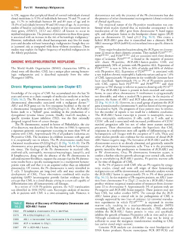
TABLE History of Discovery of Philadelphia Chromosome and tion experiments in which P210 BCR-ABL1 is expressed in murine
56.2 BCR-ABL1 Fusion marrow cells, resulting in a myeloproliferative disorder (MPD)
resembling CML, and (b) the fact that imatinib, a TKI, selectively
1960 Philadelphia chromosome (Ph) is identified. inhibits the BCR-ABL1 fusion protein in mice and specifically
1973 Ph is t(9;22)(q34;q11.2). inhibits the growth of human Ph-positive cells in vitro and in vivo.
Although considered necessary, BCR-ABL1 may not be initial or
1983 ABL1 is translocated from chromosome 9 to chromosome 22.
sufficient to cause the malignant transformation resulting in CML
1984 BCR is localized to 22q11. (see earlier clonal origin section).
1987 Ph′ is BCR-ABL1 fusion. Genomic PCR analysis can determine the exact breakpoints of
DNA fusion products. Reverse transcriptase PCR (RT-PCR) and