
Page 944 - Hematology_ Basic Principles and Practice ( PDFDrive )
P. 944
Chapter 56 Conventional and Molecular Cytogenomic Basis of Hematologic Malignancies 827
mutations followed by clonal evolution proceeding with TP53 and MULTIPLE MYELOMA
BIRC3 genomic lesions. The second evolutionary pathways involves
del(13q) and proceeds towards acquisition of SF3B1 mutations and MM is a malignancy of terminally differentiated B cells (see Chapter
BIRC3 abnormalities. These molecular events confirmed earlier 86). MM plasma cells have a very low proliferation rate, a character-
cytogenetic observations, that trisomy 12 coexists with 13q rearrange- istic that has limited the field of cytogenetic studies. Conventional
ments in rare patients. Recent integration of cytogenetic and muta- karyotyping reveals chromosomal abnormalities in 25% to 30% of
tion studies in 637 patients with newly diagnosed CLL consistently newly diagnosed patients, especially in cases with an exceptionally
showed that patients with TP53 or BIRC3 lesions had a worse high plasma cell proliferative rate. Karyotypes obtained from these
prognosis, followed by patients with mutations in SFRB1 and cells usually are complex and exhibit more than 20 aberrations in
NOTCH1 and del(11q). The presence of a minor subclone composed approximately 10% of cases.
of as little as 2% of the tumor cells within a population in early CLL When reassessed by FISH, CGH, and multicolor karyotyping
predated its emergence as a dominant subclone at relapse and the using a large panel of centromere-specific and translocation-specific
development of a chemoresistant phenotype. probes, interphase plasma cell nuclei have been shown to be charac-
terized by chromosomal aneuploidy in almost all patients with MM
or monoclonal gammopathy of unknown significance (MGUS).
Richter Syndrome Array CGH indicates that 100% of patients with newly diagnosed
MM have copy number alterations and frequent homozygous dele-
Approximately 15% of patients with CLL transform eventually to tions of genes including TRAF3, BIRC1/BIRC2, RB1 and CDKN2C.
Richter syndrome (RS), a highly aggressive phase of CLL, which Even during CR, after therapy, 12% to 71% of plasma cells still have
morphologically mimics diffuse large B-cell lymphoma (DLBCL) numerical gain or loss of chromosomes 3, 7, 8, 9, 11, 13, 15, 21,
and frequently has a dismal outcome. RS is characterized by a and X.
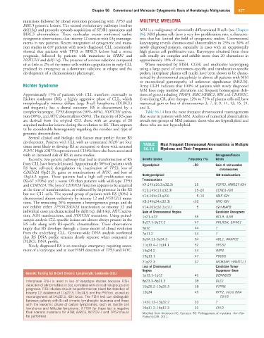
complex karyotype, TP53 disruption (50%–60%), NOTCH1 activa- Table 56.14 lists the most frequent chromosomal rearrangements
tion (30%), and MYC abnormalities (30%). The majority of RS cases that occur in patients with MM. Analysis of numerical abnormalities
are derived from the original CLL clone with an average of 20 reveals two groups of MM patients: those who are hyperdiploid and
acquired molecular lesions during the evolution to RS. There appears those who are not hyperdiploid.
to be considerable heterogeneity regarding the number and type of
genomic abnormalities.
Several clinical and biologic risk factors may predict future RS
development. Patients with CLL with an unmutated IGHV are four TABLE Most Frequent Chromosomal Abnormalities in Multiple
times more likely to develop RS as compared to those with mutated 56.14 Myeloma and Their Frequencies
IGHV. High ZAP70 expression and CD49d have also been associated
with an increased risk of RS in some studies. Oncogenes/Fusion
Recently, two genetic pathways that lead to transformation of RS Genetic Lesion Frequency (%) Genes
from CLL have been delineated. Approximately 50% of patients with Hyperdiploid ~50 Gain of odd-number
RS have cell-cycle deregulation via inactivation of TP53, loss of chromosomes
CDKN2A (9p21.3), gains or translocations of MYC, and loss of
13q14.3 region. These patients had a high cell proliferation rate Nonhyperdiploid IGH translocations
(Ki-67 >70%) and a worse OS than patients with wild-type TP53 Translocations
and CDKN2A. The loss of CDKN2A function appears to be acquired t(4;14)(p16.3;q32.3) 15 FGFR3, MMSET-IGH
at the time of transformation, as evidenced by its presence in the RS t(11;14)(q13;q32.3) 15–20 CCND1-IGH
but not CLL cells. The second group of patients with RS (30%) is t(14;16)(q13;q23) 5–10 MAF-IGH
characterized almost exclusively by trisomy 12 and NOTCH1 muta-
tions. The remaining 20% represent a heterogeneous group, and do t(8;14)(q24;q32.3) <10 MYC-IGH
not exhibit either TP53/CDKN2A inactivation or trisomy 12 and t(14;20)(q32.3;q11) 5 IGH-MAFB
individual cases are characterized by del(11q), del(14q), MYC activa- Gain of Chromosomal Region Candidate Oncogenes
tion, IGH translocations, and NOTCH1 mutations. Using paired- 1q21–q22 55 BCL9, IL6R
sample analysis CLL-specific lesions are almost always present in the
RS cells along with RS-specific abnormalities. These observations 3q27.1–3q27.2 47 POLR2H, EIF4G1
imply that RS develops through a linear model of clonal evolution 5p12 44 ?
from the underlying CLL. Genome-wide DNA analysis confirmed 7p11.2 44 ?
that RS DNA profile remains clearly separate when compared to
DLBCL DNA profile. 9q34.11–9q34.3 54 ABL1, ANAPC2
Newly diagnosed RS is an oncologic emergency requiring assess- 11q13.4–11q14.1 52 SPCS2
ment of a karyotype and at least FISH detection of TP53 and MYC. 15q24.2 44 IMP3
19q13.1 47 PDCD5
21q22.3 37 MCM3AP, HRMT1L1
Loss of Chromosomal Candidate Tumor
Region Suppressor Gene
Genetic Testing for B-Cell Chronic Lymphocytic Leukemia (CLL)
1p13.1–1p12 41 DENND2D
Interphase FISH is used in lieu of karyotype studies because FISH 8p23.3–8p21.3 28 DLC1
detection of abnormalities in CLL correlates with clinical risk groups and 10q26,2–10q26.3 18 PTPRE
prognosis. FISH studies should be performed on blood for detection of
trisomy 12, deletions of 11q22.3, 13q14.3, and the P53 loci, as well as 13q34 49 RFP2, micro RNA
rearrangement of 14q32.3, IGH locus. The FISH test can distinguish 15/16
between patients with B-cell chronic lymphocytic leukemia and those 1432.13–13q32.2 33 ?
with the leukemic phase of certain lymphomas, such as mantle cell
lymphoma and follicular lymphoma. If FISH for these loci is negative 16q11.2–16q12.3 31 CYLD
then somatic mutations for ATM, BIRC3, NOTCH 1 and TP53 should Modified from Anderson KC, Carrasco RD: Pathogenesis of myeloma. Ann Rev
be performed. Pathol 6:249, 2011.