
Page 945 - Hematology_ Basic Principles and Practice ( PDFDrive )
P. 945
828 Part VII Hematologic Malignancies
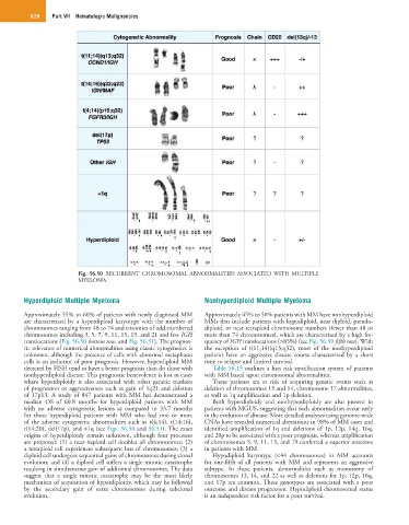
Fig. 56.50 RECURRENT CHROMOSOMAL ABNORMALITIES ASSOCIATED WITH MULTIPLE
MYELOMA.
Hyperdiploid Multiple Myeloma Nonhyperdiploid Multiple Myeloma
Approximately 55% to 60% of patients with newly diagnosed MM Approximately 45% to 50% patients with MM have nonhyperdiploid
are characterized by a hyperdiploid karyotype with the number of MMs that include patients with hypodiploid, near diploid, pseudo-
chromosomes ranging from 48 to 74 and trisomies of odd-numbered diploid, or near-tetraploid chromosome numbers (fewer than 48 or
chromosomes including 3, 5, 7, 9, 11, 15, 19, and 21 and few IGH more than 74 chromosomes), which are characterized by a high fre-
translocations (Fig. 56.50 bottom row, and Fig. 56.51). The prognos- quency of IGH translocations (>85%) (see Fig. 56.50 fifth row). With
tic relevance of numerical abnormalities using classic cytogenetics is the exception of t(11;14)(q13;q32), most of the nonhyperdiploid
unknown, although the presence of cells with abnormal metaphases patients have an aggressive disease course characterized by a short
cells is an indicator of poor prognosis. However, hyperdiploid MM time to relapse and limited survival.
detected by FISH tend to have a better prognosis than do those with Table 56.15 outlines a lists risk stratification system of patients
nonhyperdiploid disease. This prognostic benevolence is lost in cases with MM based upon chromosomal abnormalities.
where hyperdiploidy is also associated with other genetic markers These patients are at risk of acquiring genetic events such as
of progression or aggressiveness such as gain of 1q21 and deletion deletion of chromosomes 13 and 14, chromosome 17 abnormalities,
of 17p13. A study of 847 patients with MM has demonstrated a as well as 1q amplification and 1p deletion.
median OS of 60.8 months for hyperdiploid patients with MM Both hyperdiploidy and nonhyperdiploidy are also present in
with no adverse cytogenetic lesions as compared to 33.7 months patients with MGUS, suggesting that such abnormalities occur early
for those hyperdiploid patients with MM who had one or more in the evolution of disease. More detailed analyses using genome-wide
of the adverse cytogenetic abnormalities such as t(4;14), t(14;16), CNAs have revealed numerical aberrations in 98% of MM cases and
t(14;20), del(17p), and +1q (see Figs. 56.50 and 56.51). The exact identified amplification of 1q and deletions of 1p, 12p, 14q, 16q,
origins of hyperdiploidy remain unknown, although four processes and 20p to be associated with a poor prognosis, whereas amplification
are proposed: (1) a near-haploid cell doubles all chromosomes; (2) of chromosomes 5, 9, 11, 15, and 19 conferred a superior outcome
a tetraploid cell experiences subsequent loss of chromosomes; (3) a in patients with MM.
diploid cell undergoes sequential gains of chromosomes during clonal Hypodiploid karyotype (<44 chromosomes) in MM accounts
evolution; and (4) a diploid cell suffers a single mitotic catastrophe for one-fifth of all patients with MM and represents an aggressive
resulting in simultaneous gain of additional chromosomes. The data subtype. In these patients, abnormalities such as monosomy of
suggest that a single mitotic catastrophe may be the most likely chromosomes 13, 14, and 22 as well as deletions for 1p, 12p, 16q,
mechanism of acquisition of hyperdiploidy, which may be followed and 17p are common. These genotypes are associated with a poor
by the secondary gain of extra chromosomes during subclonal outcome and disease progression. Hypodiploid chromosomal status
evolution. is an independent risk factor for a poor survival.