
Page 1078 - Williams Hematology ( PDFDrive )
P. 1078
1052 Part VIII: Monocytes and Macrophages Chapter 67: Structure, Receptors, and Functions of Monocytes and Macrophages 1053
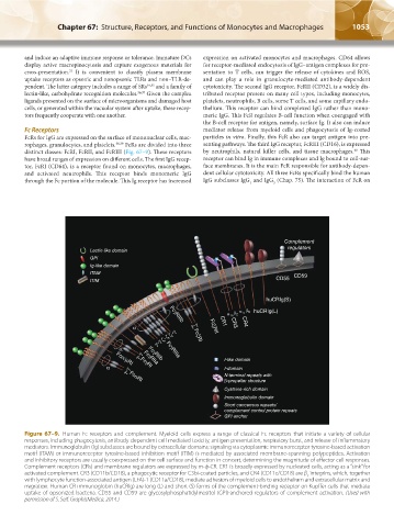
and induce an adaptive immune response or tolerance. Immature DCs expression on activated monocytes and macrophages. CD64 allows
display active macropinocytosis and capture exogenous materials for for receptor-mediated endocytosis of IgG–antigen complexes for pre-
33
cross-presentation. It is convenient to classify plasma membrane sentation to T cells, can trigger the release of cytokines and ROS,
uptake receptors as opsonic and nonopsonic TLRs and non–TLR-de- and can play a role in granulocyte-mediated antibody-dependent
pendent. The latter category includes a range of SRs 34,35 and a family of cytotoxicity. The second IgG receptor, FcRII (CD32), is a widely dis-
lectin-like, carbohydrate recognition molecules. 36,37 Given the complex tributed receptor present on many cell types, including monocytes,
ligands presented on the surface of microorganisms and damaged host platelets, neutrophils, B cells, some T cells, and some capillary endo-
cells, or generated within the vacuolar system after uptake, these recep- thelium. This receptor can bind complexed IgG rather than mono-
tors frequently cooperate with one another. meric IgG. This FcR regulates B-cell function when coengaged with
the B-cell receptor for antigen, namely, surface Ig. It also can induce
Fc Receptors mediator release from myeloid cells and phagocytosis of Ig-coated
FcRs for IgG are expressed on the surface of mononuclear cells, mac- particles in vitro. Finally, this FcR also can target antigen into pre-
rophages, granulocytes, and platelets. 38,39 FcRs are divided into three senting pathways. The third IgG receptor, FcRIII (CD16), is expressed
40
distinct classes: FcRI, FcRII, and FcRIII (Fig. 67–9). These receptors by neutrophils, natural killer cells, and tissue macrophages. This
have broad ranges of expression on different cells. The first IgG recep- receptor can bind Ig in immune complexes and Ig bound to cell-sur-
tor, FcRI (CD64), is a receptor found on monocytes, macrophages, face membranes. It is the main FcR responsible for antibody-depen-
and activated neutrophils. This receptor binds monomeric IgG dent cellular cytotoxicity. All three FcRs specifically bind the human
through the Fc portion of the molecule. This Ig receptor has increased IgG subclasses IgG and IgG (Chap. 75). The interaction of FcR on
1
3
Complement
regulators
Lectin-like domain
GPI
Ig-like domain
ITAM CD59
CD55
ITIM
huCRIg(S)
α m β 2 α x β 2 huCRIg(L)
FcγRIIIb
α ζ–γ γ–γ FcξRII CR1 CR3 CR4
γ–γ ζ–ζ FcξRI
α FcγRIIIa
γ–γ FcγRIIa I-like domain
FcγRIIb
Fcα/µRI
α FcγRI I-domain
γ–γ N-terminal repeats with
β-propeller structure
FcαRI
Cysteine-rich domain
Immunoglobulin domain
Short concensus repeats/
complement control protein repeats
GPI anchor
Figure 67–9. Human Fc receptors and complement. Myeloid cells express a range of classical Fc receptors that initiate a variety of cellular
responses, including phagocytosis, antibody-dependent cell-mediated toxicity, antigen presentation, respiratory burst, and release of inflammatory
mediators. Immunoglobulin (Ig) subclasses are bound by extracellular domains; signaling via cytoplasmic immunoreceptor tyrosine-based activation
motif (ITAM) or immunoreceptor tyrosine-based inhibition motif (ITIM) is mediated by associated membrane-spanning polypeptides. Activation
and inhibitory receptors are usually coexpressed on the cell surface and function in concert, determining the magnitude of effector cell responses.
Complement receptors (CRs) and membrane regulators are expressed by m-ф-CR. CR1 is broadly expressed by nucleated cells, acting as a “sink” for
activated complement. CR3 (CD11b/CD18), a phagocytic receptor for C3bi-coated particles, and CR4 (CD11c/CD18) are β integrins, which, together
2
with lymphocyte function-associated antigen (LFA)-1 (CD11a/CD18), mediate adhesion of myeloid cells to endothelium and extracellular matrix and
migration. Human CR immunoglobin (huCRIg) are long (L) and short (S) forms of the complement-binding receptor on Kupffer cells that mediate
uptake of opsonized bacteria. CD55 and CD59 are glycosylphosphatidylinositol (GPI)-anchored regulators of complement activation. (Used with
permission of S. Seif, GraphisMedica, 2014.)
Kaushansky_chapter 67_p1043-1074.indd 1053 9/21/15 10:43 AM