
Page 1085 - Williams Hematology ( PDFDrive )
P. 1085
1060 Part VIII: Monocytes and Macrophages Chapter 67: Structure, Receptors, and Functions of Monocytes and Macrophages 1061
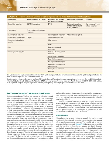
TABLE 67–5. Selected G-Protein–Coupled Receptors Implicated in Functions of Monocytes and Macrophages
Chemotaxis Adhesion/Cell–Cell Contact Activation and Resolu- Alternative Activation Survival
tion of Inflammation
Chemokine receptors EGF-TM7 receptors BAI-1 Purinergic receptors Sphingosine-1-phos-
GPR86, GPR105, P2Y8, phate receptors
P2Y11, and P2Y12
C5a receptor Sphingosine-1-phosphate
receptors
Leukotriene B receptor Formyl peptide receptors Chemokine receptors
4
Formyl peptide receptors CX CR1 Chemokine receptors
3
Platelet-activating factor C5a receptor
receptor
EMR2
EMR2 Protease-activated
receptors
Neuropeptide Y receptor Platelet-activating factor
receptor
Leukotriene B receptor
4
Neurokinin receptors
Neuropeptide Y receptor
Vasoactive intestine pep-
tide receptor
Prostaglandin receptors
Resolvin
BAI-1, brain-specific angiogenesis inhibitor 1; EGF-TM7, epidermal growth factor–seven transmembrane; EMR2, epidermal growth factor–like
module containing mucin-like hormone receptor–like 2.
Data from Lattin, J.E. et al.: Expression analysis of G Protein-Coupled Receptors in mouse macrophages. Immunome Res 4:5, 2008; Yona, S., Lin,
H.H., Siu, W.O., Gordon, S. & Stacey, M.: Adhesion-GPCRs: emerging roles for novel receptors. Trends Biochem Sci 33:491-500, 2008; Lattin, J.
et al.: G-protein-coupled receptor expression, function, and signaling in macrophages. J Leukoc Biol 82:16–32, 2007.
RECOGNITION AND CLEARANCE OVERVIEW and engulfment of erythrocytes can be visualized by scanning elec-
Resident macrophages of the liver and marrow, as well as in lung and tron microscopy, and the sequence of engulfment by phase-contrast
other nonhematopoietic tissues, play a major role in the recognition, optics (see video talk on macrophage phagocytosis at http://hstalks.
phagocytosis, and endocytosis of foreign particles and macromole- com/?t=BL1473311).
cules, as well as of modified host components. Clearance can be silent, In addition, interest has grown explosively in cytosolic recognition
even suppressing inflammation, mediated by transforming growth systems, designed to protect the cell from various infectious and lytic
100–102
factor (TGF)-β generation, as observed after the uptake of apoptotic agents. The process of autophagy shares aspects with both mem-
cells by macrophages. Production of hematopoietic cells is balanced brane-bound and cytoplasmic organelle injury, and has become of great
99
by their programmed senescence and increased destruction, which current interest because of its contribution to pathogenesis of infec-
103
can be enhanced in response to microbial and other toxic substances. tious, malignant, and inflammatory syndromes.
Macrophages initiate and perpetuate inflammation, both acute and
chronic, as a result of their biosynthetic and secretory responses to APOPTOSIS
injurious particles. Uptake and vacuole formation sequester the mem- Macrophages take up large numbers of naturally dying cells, hemato-
brane-enclosed contents for digestion and possible antigen processing poietic and others, through a complex mechanism involving multiple,
and presentation, a specialized property of DCs after their further dif- often redundant nonopsonic receptors. 47,99 A possible role for comple-
ferentiation from active endocytic to APCs. Specialized studies show ment has also been proposed. Figure 67–12 illustrates receptors and lig-
33
that blood-derived monocytes have unique functions. For example, ands that have been implicated. Apart from the SRs already discussed,
in the human disorder multiple sclerosis and the model experimen- they include receptors for opsonins and for milk-fat globulin, as well as
tal autoimmune encephalitis, monocyte-derived macrophages initiate for the vitronectin receptor. Phosphatidylserine (PS) expressed on the
demyelination at nodes of Ranvier; whereas, microglia derived from outer leaflet of apoptotic cells, contributes to apoptotic cell recognition,
yolk-sac progenitors during embryogenesis are relatively inert at dis- but its role is probably more complex as apparently healthy cells can
ease onset. To illustrate the role of macrophages in the recognition express patches of PS on their surface and PS recognition plays a role in
31
and clearance of foreign substances, images of macrophage spreading CD36-dependent macrophage–macrophage fusion. The recognition
104
Kaushansky_chapter 67_p1043-1074.indd 1060 9/21/15 10:43 AM