
Page 1087 - Williams Hematology ( PDFDrive )
P. 1087
1062 Part VIII: Monocytes and Macrophages Chapter 67: Structure, Receptors, and Functions of Monocytes and Macrophages 1063
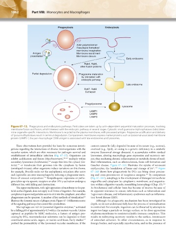
Phagocytosis Endocytosis
Actin polymerization
Pseudopod formation/
membrane invagination
Antigen Membrane recruitment
presentation Membrane closure
Early endosome
Rab4, Rab5,
other fusion proteins
Partial degradation Phagosome maturation
by interaction with
endocytic pathway
Late endosome
Rab7, Rab9
Cathepsin
Phagolysosome
Lysosome
LAMP1
LAMP2
LAMP3
Figure 67–13. Phagocytosis and endocytosis pathways. Particulates are taken up by actin-dependent sequential maturation processes, involving
membrane fusion and fission, which intersect with the endocytic pathway at several stages. Cytosolic small guanosine triphosphatases (rabs) deter-
mine organelle-specific interactions. Membrane is recycled to the plasma membrane, with processed antigen. Progressive acidification and delivery
of lysosomal hydrolases result in terminal degradation. Compartment membranes express marker proteins such as lysosomal-associated membrane
protein (LAMP)-1; the pan-macrophage CD68 antigen is associated with late endosomes and lysosomes.
These observations have provided the basis for numerous investi- content cannot be fully degraded because of its nature (e.g., sucrose),
gations regarding the interactions of diverse microorganisms with the overload (e.g., lipid), or owing to a genetic deficiency in a catabolic
vacuolar system, which are often necessary for pathogen survival and enzyme (lysosomal storage diseases), it accumulates within residual
establishment of intracellular infection (Fig. 67–15). Organisms can lysosomes, altering macrophage gene expression and secretory out-
inhibit acidification and fusion (Mycobacterium), 101,113 multiply within put, thus mediating chronic inflammation or metabolic forms of mod-
secondary lysosomes (Leishmania), escape free into the cytosol (Lis- ified inflammation, such as atherosclerosis, foam cell formation and
114
teria), or translocate their genomes into the cytoplasm by fusion Gaucher disease. Figure 67–16A illustrates the uptake of senescent
115
(enveloped viruses); other organisms induce variations on this theme; erythrocytes, the breakdown of heme and storage of Fe . Figure
2+ 118
for example, Brucella seeks out the endoplasmic reticulum after entry 67–16B shows how phagocytosis by DCs can bring about process-
and Legionella can enter macrophages by inducing a phagosome mem- ing and cross-presentation of exogenous antigens. By comparison
119
brane of unusual composition. Nonpathogenic organisms or patho- (Fig. 67–16C), autophagy is the envelopment of damaged intracellular
116
gens taken up via opsonic receptors or after IFN-γ activation undergo a organelles and cytoplasm by cytoplasmic membrane, and sequestra-
different fate, with killing and destruction. tion within a digestive vacuole, resembling heterophagy (Chap. 15).
116
The zipper mechanism, with tight apposition of membrane to the par- Its biochemical and cellular basis has become of interest because of
ticle’s surface ligands, does not apply to all forms of ingestion. For example, its apparent relevance to cancer, infections such as tuberculosis and
complement opsonized particles seem to sink into the cytoplasm, and other Legionnaire disease, and inflammatory syndromes such as inflamma-
phagosomes can be spacious. A number of key methods of visualization tory bowel disease (IBD).
109
illustrate the dynamic nature of phagocytosis. Figure 67–14 illustrates some Although the phagocytic mechanism has been investigated in
of the signaling pathways that control the cytoskeleton. depth, we do not understand fully how the process of internalization
Macrophages are rich in lysosomal digestive enzymes, activated is controlled. For example, ingestion can be thwarted by attempts to
33
by a falling pH of approximately 6.5 within the mature vacuole. Unless ingest too large a particle or foreign surface, or by close apposition
captured as peptides by MHC molecules, a feature of antigen pro- of plasma membrane to noninternalizable immune complexes. This
cessing by DCs, macromolecular substrates can be degraded to their results in redirecting secretory vesicles to the surface, reminiscent
constituent amino acids, sugars, or nucleic acid bases. Early studies of osteoclast adhesion. In other circumstances, as in response to
117
probed the permeability of the lysosomal vacuolar membrane. If the foreign bodies, and especially mycobacteria, and in the presence of
Kaushansky_chapter 67_p1043-1074.indd 1062 9/21/15 10:43 AM