
Page 1086 - Williams Hematology ( PDFDrive )
P. 1086
1060 Part VIII: Monocytes and Macrophages Chapter 67: Structure, Receptors, and Functions of Monocytes and Macrophages 1061
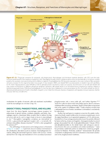
Figure 67–12. Phagocytic receptors for apoptotic cell phagocytosis. Macrophages and immature myeloid dendritic cells (DCs) are the main
immune cells involved in the clearance of apoptotic cells. They express broadly similar multiple receptors that can bind directly or via opsonic-soluble
proteins, for example, mannose-binding lectins (MBLs) to ligands. Phosphatidylserine (PS) becomes exposed on the outer surface of the apoptotic
cell and a receptor for this ligand has been long sought. A new receptor (TIM4, and related TIM1) was discovered on resident mф, with specificity
for PS. Other mф populations utilize MFGE8 (a milk-fat globulin protein secreted by mф) as an opsonin. Discrimination of non-self and altered self
may involve combinations of different phagocyte receptors. Apoptotic cell uptake results in an antiinflammatory response by mф (e.g., release of
transforming growth factor [TGF]-β and prostaglandin E ), but has also been implicated in cross-presentation by DCs. For further details see Ref. 47.
2
(Reproduced with permission of Savill J, Dransfield I, Gregory C, et al: A blast from the past: clearance of apoptotic cells regulates immune responses. Nat Rev
Immunol 2002 Dec;2(12):965-975.)
mechanisms for uptake of necrotic cells and enucleated erythroblast phagolysosomes with a more acidic pH, and further digestion. 108,109
nuclei by macrophages are not clear (Chap. 15). Apart from selective fusion with intracellular vesicles, there is extensive
membrane flow, recycling, and fusion. Small GTPases play an important
role in the control of membrane traffic. Early estimates revealed that
110
ENDOCYTOSIS, PHAGOCYTOSIS, AND KILLING a substantial fraction of surface membrane is internalized constitutively
Apart from the above ligands, macrophages express receptors for by endocytosis.
endocytosis of growth factors, cytokines, peptides, and lipids. Mac- Studies that used opsonic receptors to examine the uptake mecha-
rophages express a functional folate receptor that is induced during nism of antibody-coated erythrocytes via opsonic receptors gave rise to
activation and can be used to target drugs or tracers to macrophages the zipper hypothesis: local segmental engagement of FcR, and circum-
in situ. Hemoglobin–haptoglobin complexes are internalized by ferential flow of macrophage pseudopodia around the particle, followed
105
CD163, a glucocorticoid-regulated receptor with a remarkable SR- by fusion at the tip, closure, and ingestion. Subsequent studies by several
cysteine extracellular domain structure. CD163 is also upregulated groups documented the role of phosphatidylinositol 3-kinase (PI3K)
106
by substance P. 107 and phosphoinositides in the initial fusion and subsequent associa-
The cell biology of endocytosis and of phagocytosis is illustrated tions between the actin cytoskeleton and cellular membranes. Latex
111
in Figs. 67–13 and 67–14. Apart from size and resultant involvement of has provided a useful test particle to isolate latex-containing phagoly-
the cytoskeleton, they have much in common; vesicle/phagosome for- sosomes by flotation. Proteomic analyses demonstrated the protein
112
mation, falling pH and initial digestion, fusion with secretory vesicles composition of phagosomes and drew attention to functional constitu-
derived from the Golgi, and maturation to form secondary lysosomes/ ents in the phagolysosomal membrane.
Kaushansky_chapter 67_p1043-1074.indd 1061 9/21/15 10:43 AM