
Page 1088 - Williams Hematology ( PDFDrive )
P. 1088
1062 Part VIII: Monocytes and Macrophages Chapter 67: Structure, Receptors, and Functions of Monocytes and Macrophages 1063
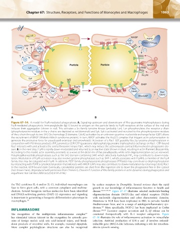
Figure 67–14. A model for FcγR-mediated phagocytosis. A. Signaling upstream and downstream of Rho guanosine triphosphatases during
FcγR-mediated phagocytosis. Immunoglobulin (Ig) G bound to antigen on the particle binds to FcγRI receptors at the surface of the mф and
induces their aggregation (shown in red). This activates a Src family tyrosine kinase (probably Lyn). Lyn phosphorylates the receptor γ chain
(phosphotyrosine residues in the γ chains are depicted as red diamonds) and Syk. Syk is activated and recruited to the phosphotyrosine residues
of the γ chain through its two SH2 (Src homology 2) domains. Cdc42 activation by an unknown guanine–nucleotide exchange factor (GEF) allows
the recruitment of WASP (Wiskott-Aldrich syndrome protein). In turn, WASP activates the Arp2/3 complex that triggers actin polymerization to
generate the protrusive force for pseudopod extension (red arrowheads). Activation of a Rac1 GEF, possibly Vav, by tyrosine phosphorylation in
conjunction with PI3 kinase products (PIP ) promotes GDP/GTP (guanosine diphosphate/guanosine triphosphate) exchange on Rac1. GTP-bound
3
Rac1 interacts with and activates the serine/threonine kinase Pak1, which may induce the actinomyosin contractility involved in phagosome clo-
sure. B. In the next step, FcγRI is rapidly down-modulated and returned to an inactive state (shown in blue), resulting in actin filament disassembly.
According to this model, actin assembly proceeds as a wave at the distal rim of the pseudopodia, while actin depolymerization occurs rearward.
Polyphosphoinositide phosphatases such as the SH2 domain-containing SHIP, which selectively hydrolyze PIP , may contribute to down-modu-
3
lation. Modulation of FcγRI activation may also involve tyrosine phosphatases such as SHP-1, which associates with FcγRIIb, a member of the FcγR
family that may be coligated with FcγRI. In addition, PEST family phosphotyrosine phosphatases (PTPases) may contribute to dephosphorylation
by interacting with PSPIP, a cytoskeletal protein that interacts with WASP. GAPs may also contribute to down-modulation by returning Cdc42/Rac1
to the inactive, GDP-bound state. Eventually, cytoskeletal proteins are shed from the ingestion site to leave the phagosome free in the cytosol
(not shown here). (Reproduced with permission from Chimini G, Chavrier P: Function of Rho family proteins in actin dynamics during phagocytosis and
engulfment. Nat Cell Biol 2000 Oct;2(10):E191-E196.)
the Th2 cytokines IL-4 and/or IL-13, individual macrophages can by surface receptors in Drosophila. Several reviews chart the rapid
fuse to form giant cells, with a common cytoplasm and multinu- growth in our knowledge of inflammasome function in health and
cleation. Several fusogenic surface molecules have been identified disease. 100,102,121,122 Figure 67–17 illustrates selected nucleotide-binding
and DNAX-activating protein (DAP) 12 expression and signaling oligomerization domain (NOD)-like and related receptors (NLRs)
is important in generating a fusogenic differentiation phenotype in with nucleotide oligomerization and other characteristic domains.
macrophages. 120 Mutations in NLR have been implicated in IBD, in periodic familial
Mediterranean fever, and in a range of autohyperinflammatory syn-
dromes. More specifically, NOD-2 has been implicated in Crohn
123
INFLAMMASOME disease. 124,125 Excessive caspase activation and IL-1β release can be
101
The recognition of the multiprotein inflammasome complex countered therapeutically with IL-1 receptor antagonists. Figure
has stimulated intense interest in the recognition by cytosolic pro- 67–18 illustrates the role of inflammasome activation in intracellular
teins of foreign nucleic acid, uric acid-induced injury, and break- infection. Antiviral production of IFN-α and -β involves retinoid-
down products of microbial walls, for example, muramyl dipeptide. inducible gene (RIG)-I–like helicases, indicating a role for mitochon-
More complex peptidoglycan structures can also be recognized dria in cytosolic sensing.
Kaushansky_chapter 67_p1043-1074.indd 1063 9/21/15 10:43 AM