
Page 1950 - Williams Hematology ( PDFDrive )
P. 1950
1924 Part XII: Hemostasis and Thrombosis Chapter 113: Molecular Biology and Biochemistry of the Coagulation Factors 1925
2 4 6 8 10 12 14 22 24 FACTOR VIII
1 3 5 7 9 11 13 15–1718–21 23 25
Gene 70 kb Factor VIII (antihemophilic factor) was first discovered in 1937, but it
was not until 1979 that its purification by Tuddenham and coworkers
led to the molecular identification of the protein. 139,140 Factor VIII is
synthesized as a single-chain preprocofactor of 2351 amino acids and,
mRNA 7 kb subsequent to intracellular processing, is secreted as a series of metal
ion-linked heterodimers due to proteolysis at the A3-B junction and dif-
ferential processing in the central B domain (Fig. 113–13). The mature
factor VIII procofactor comprises 2332 amino acids (Mr ≈300,000) and
circulates in a high-affinity complex with its carrier protein VWF at a
Exon 12 34 56 78 10 12 13 1516 18 212223 25 concentration of approximately 0.7 nM and a circulatory half-life of 8
Protein P A1 A2 B A3 C1C2
to 12 hours (see Table 113–1). Complex formation with VWF protects
Figure 113–12. Relationship of gene structure to protein structure factor VIII from proteolytic degradation, premature ligand binding, and
in factor V. The exons, introns, mRNA, and protein structure are as indi- rapid clearance from the circulation.
cated. The mRNA is 7 kb with some 5′ and 3′ untranslated sequences The primary source of factor VIII is the liver, 141,142 but extrahepatic
(light blue). In the protein, P indicates the propeptide leader sequence, synthesis of factor VIII also occurs. 143,144 While contradictory evidence
and the A1-A2-B-A3-C1-C2 domains are indicated.
exists on the cellular origin of both hepatic and extrahepatic factor VIII
synthesis, recent studies in mice support that endothelial cells from
many tissues and vascular beds synthesize factor VIII, with a large con-
dissociates and factor Va can no longer associate with factor Xa. A tribution from hepatic sinusoidal endothelial cells. 145–147 This is consis-
130
common Arg506Gln mutation in factor V leads to resistance to inacti- tent with observations on factor VIII expression in human endothelial
vation by APC (factor V Leiden) and is associated with an increased risk cells from the liver and lung. 148,149
of venous thromboembolism (Chap. 133). 131 Factor VIII is less-efficiently secreted from the cell as compared
Both factor V and an alternatively spliced isoform of factor V to factor V, because it interacts with the ER-chaperon proteins calnexin
(factor V-short), which lacks the major part of the B domain (residues and calreticulin, whereas factor V interacts with calreticulin only.
150
756 to 1458) and normally circulates in low abundance, interact with Both chaperons preferentially interact with GPs comprising mono-
full-length TFPI (TFPIα), most likely through the acidic B domain glucosylated N-linked oligosaccharides and promote correct folding of
region. 132,133 The linkage of factor V and TFPIα is considered to atten- proteins that enter the secretory pathway and target misfolded proteins
uate the bleeding phenotype in factor V–deficient patients, as the low for degradation. Factor VIII, but not factor V, also interacts with the
TFPIα levels in these patients allow the residual platelet factor V to be ER-chaperon immunoglobulin-binding protein (BiP/GRP78), which
sufficient for coagulation. 132,134 Conversely, increased factor V–short appears to enhance the stability of factor VIII, but also retards its secre-
expression caused by an A2440G mutation in the factor V gene leads to tion. Factor VIII trafficking from the ER to the Golgi is mediated via
151
a dramatic increase in plasma TFPIα, resulting in a bleeding disorder. 133 the LMAN1-MCDF2 receptor complex, similar to factor V. 103
Several clearance receptors are responsible for actively removing fac-
Gene Structure and Variations tor VIII from the circulation, which include the low-density lipoprotein
The gene for factor V (F5) is located on chromosome 1q23. It is located (LDL) receptor-related protein 1 (LRP1), the LDL receptor, and receptors
very close to the genes for the selectin family of leukocyte adhesion that specifically interact with carbohydrate structures on factor VIII. 152–156
molecules. The factor V gene spans approximately 70 kb and consists of
25 exons (Fig. 113–12). The gene structure is very similar to that of the Protein Structure
factor VIII gene, with exon–intron boundaries occurring at exactly the The A1-A2-B-A3-C1-C2 domain structure of factor VIII shares
same location in 21 out of 24 cases. 135 significant homology with factor V except in the B domain region
Homozygosity or compound heterozygosity for loss-of-function (see Fig. 113–13). In contrast to factor V, the factor VIII B domain is
mutations in the factor V gene lead to a bleeding disorder (termed par- dispensable for procoagulant activity. The mature factor VIII proco-
ahemophilia or Owren parahemophilia). At the time of writing, 152 factor comprises a variably sized heavy chain (A1-A2-B; Mr ≈200,000
136
mutations in the factor V gene have been collected in the human gene to 90,000 depending on the extent of proteolysis) and a light chain
mutation database (www.hgmd.org). (A3-C1-C2; Mr ≈80,000). The C-terminal regions of the A1 and A2
Gain-of-function mutations in the factor V gene increase the domains and the N-terminal portion of the A3 domain contain short
risk of thrombosis. This is particularly the case for venous thrombosis segments of 30 to 40 negatively charged residues known as the a1, a2,
and not so much for arterial thrombosis. In whites, the most common and a3 regions. Interaction with VWF is facilitated by the a3 region and
gain-of-function mutation in the factor V gene is factor V Leiden (Arg- C1 domain. 157,158 The C domains mediate binding to the anionic phos-
506Gln), which leads to a plasma abnormality known as APC resistance pholipid surface, thereby localizing factor VIII to the site of injury and
(Chap. 133). 137,138 facilitating interaction with factor IXa and factor X. 159–161
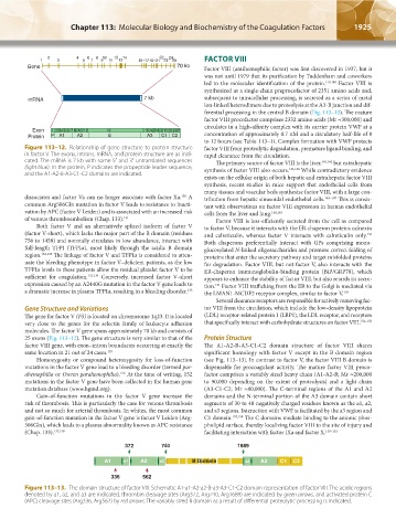
372 740 1689
A1 a1 A2 a2 B Domain a3 A3 C1 C2
336 562
Figure 113–13. The domain structure of factor VIII. Schematic A1-a1-A2-a2-B-a3-A3-C1-C2 domain representation of factor VIII. The acidic regions
denoted by a1, a2, and a3 are indicated, thrombin cleavage sites (Arg372, Arg740, Arg1689) are indicated by green arrows, and activated protein C
(APC) cleavage sites (Arg336, Arg562) by red arrows. The variably sized B domain as a result of differential proteolytic processing is indicated.
Kaushansky_chapter 113_p1915-1948.indd 1925 9/21/15 2:39 PM