
Page 1951 - Williams Hematology ( PDFDrive )
P. 1951
1926 Part XII: Hemostasis and Thrombosis Chapter 113: Molecular Biology and Biochemistry of the Coagulation Factors 1927
Factor VIII is heavily glycosylated and the majority of the N-linked THE SOLUBLE COFACTORS PROTEIN S
glycosylation sites are found in the B domain, which mediate interac-
tion with the chaperons calnexin and calreticulin and, in part, with the AND VON WILLEBRAND FACTOR
LMAN1–MCDF2 receptor complex. 103,150,162 Sulfation of tyrosine resi-
dues is required for optimal activation by thrombin, maximal activity PROTEIN S
in complex with factor IXa, and maximal affinity of factor VIIIa for Protein S, which is named after the city (Seattle) where it was discov-
VWF. 35,163 Factor VIII comprises two phosphorylation sites that are ered by the group of Earl Davie in 1977, is a vitamin K–dependent
located in the A1 (Thr351) and B (Ser1657) domains. single-chain GP of 635 amino acids (Mr ≈75,000) that circulates with a
plasma half-life of 42 hours (see Table 113–1). Part of the total protein
Factor VIII Procofactor Activation and Factor VIIIa Cofactor S pool circulates in a free form at a concentration of 150 nM, whereas
Function the majority (approximately 60 percent; 200 nM) circulates bound to
Thrombin and factor Xa are the principal activators of the procofac- the complement regulatory protein C4b–binding protein (C4BP).
tor VIII and generate the cofactor VIIIa through sequential proteoly- Protein S is primarily synthesized in the liver by hepatocytes, in addi-
sis at Arg740, Arg372, and Arg1689. 126,164–166 The heterotrimeric factor tion to endothelial cells, megakaryocytes, testicular Leydig cells, and
VIIIa is composed of the A1 (Mr ≈50,000), A2 (Mr ≈43,000), and the osteoblasts. 174–178
A3-C1-C2 light chain (Mr ≈73,000) subunits (see Fig. 113–13). The A1
and A3-C1-C2 subunits are noncovalently linked through calcium ions, Protein Structure
whereas A2 is associated with weak affinity primarily by electrostatic The protein structure of protein S differs from the other vitamin
interactions. 167,168 Once activated, factor VIIIa functions as a cofactor K–dependent proteins as it lacks a serine protease domain and, con-
for factor IXa in the phospholipid-dependent conversion of factor X to sequently, is not capable of catalytic activity. Protein S is composed of
factor Xa. The rapid and spontaneous loss of factor VIIIa cofactor activ- a Gla domain comprising 11 Gla residues, a thrombin-sensitive region
ity is attributed to A2 domain dissociation from the heterotrimer. 167,168 (TSR), four EGF domains, and a C-terminal sex hormone–binding
Additional proteolysis by APC, factor Xa, or factor IXa also results in globulin (SHBG)-like region that consists of two laminin G-type
the downregulation of factor VIIIa cofactor activity. 169 domains (Fig. 113–15). The SHBG-like domain is involved in the inter-
action with the β-subunit of C4BP.
Gene Structure and Variations Apart from γ-carboxylation of Glu residues, protein S is posttrans-
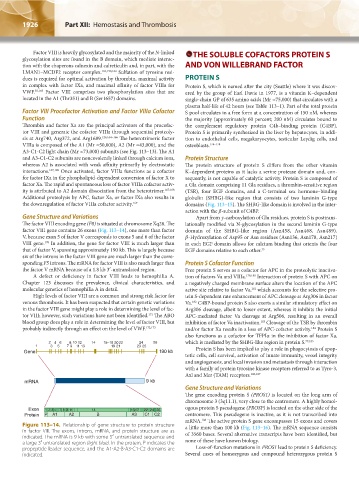
The factor VIII encoding gene (F8) is situated at chromosome Xq28. The lationally modified via N-glycosylation in the second laminin G-type
factor VIII gene contains 26 exons (Fig. 113–14), one more than factor domain of the SHBG-like region (Asn458, Asn468, Asn489).
V, because exon 5 of factor V corresponds to exons 5 and 6 of the factor β-Hydroxylation of Asp95 or Asn residues (Asn136, Asn178, Asn217)
170
VIII gene. In addition, the gene for factor VIII is much larger than in each EGF domain allows for calcium binding that orients the four
that of factor V, spanning approximately 190 kb. This is largely because EGF domains relative to each other. 35
six of the introns in the factor VIII gene are much larger than the corre-
sponding F5 introns. The mRNA for factor VIII is also much larger than Protein S Cofactor Function
the factor V mRNA because of a 1.8 kb 3′-untranslated region. Free protein S serves as a cofactor for APC in the proteolytic inactiva-
A defect or deficiency in factor VIII leads to hemophilia A. tion of factors Va and VIIIa. 179,180 Interaction of protein S with APC on
Chapter 123 discusses the prevalence, clinical characteristics, and a negatively charged membrane surface alters the location of the APC
molecular genetics of hemophilia A in detail. active site relative to factor Va, which accounts for the selective pro-
181
High levels of factor VIII are a common and strong risk factor for tein S-dependent rate enhancement of APC cleavage at Arg306 in factor
venous thrombosis. It has been suspected that certain genetic variations Va. C4BP-bound protein S also exerts a similar stimulatory effect on
182
in the factor VIII gene might play a role in determining the level of fac- Arg306 cleavage, albeit to lower extent, whereas it inhibits the initial
171
tor VIII; however, such variations have not been identified. The ABO APC-mediated factor Va cleavage at Arg506, resulting in an overall
blood group does play a role in determining the level of factor VIII, but inhibition of factor Va inactivation. Cleavage of the TSR by thrombin
183
probably indirectly through an effect on the level of VWF. 172,173 and/or factor Xa results in a loss of APC-cofactor activity. Protein S
184
also functions as a cofactor for TFPIα in the inhibition of factor Xa,
2 4 6 8 10 12 14 15–18 20 22 24 26 which is mediated by the SHBG-like region in protein S. 77,185
1 3 5 7 9 11 13 19 21 23 25 Protein S has been implied to play a role in phagocytosis of apop-
Gene 190 kb
totic cells, cell survival, activation of innate immunity, vessel integrity
and angiogenesis, and local invasion and metastasis through interaction
with a family of protein tyrosine kinase receptors referred to as Tyro-3,
Axl and Mer (TAM) receptors. 186,187
mRNA 9 kb
Gene Structure and Variations
The gene encoding protein S (PROS1) is located on the long arm of
chromosome 3 (3q11.1), very close to the centromere. A highly homol-
Exon 12 3 4 78 911 14 1617 222426 ogous protein S pseudogene (PROSP) is located on the other side of the
Protein P A1 A2 B A3 C1 C2 centromere. This pseudogene is inactive, as it is not transcribed into
mRNA. The active protein S gene encompasses 15 exons and covers
188
Figure 113–14. Relationship of gene structure to protein structure a little more than 100 kb (Fig. 113–16). The mRNA sequence consists
in factor VIII. The exons, introns, mRNA, and protein structure are as of 3560 bases. Several alternative transcripts have been identified, but
indicated. The mRNA is 9 kb with some 5′ untranslated sequence and none of these have known biology.
a large 3′ untranslated region (light blue). In the protein, P indicates the
propeptide leader sequence, and the A1-A2-B-A3-C1-C2 domains are Loss-of-function mutations in PROS1 lead to protein S deficiency.
indicated. Several cases of homozygous and compound heterozygous protein S
Kaushansky_chapter 113_p1915-1948.indd 1926 9/21/15 2:39 PM