
Page 250 - Williams Hematology ( PDFDrive )
P. 250
224 Part IV: Molecular and Cellular Hematology Chapter 16: Cell-Cycle Regulation and Hematologic Disorders 225
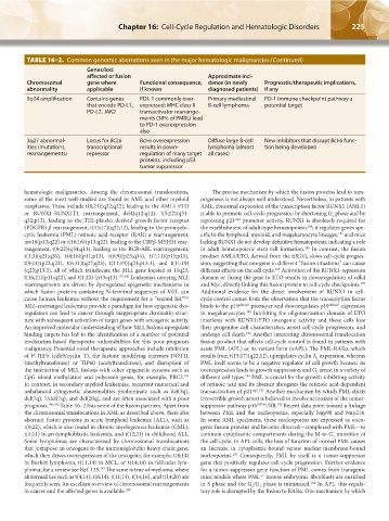
TABLE 16–2. Common genomic aberrations seen in the major hematologic malignancies.(Continued)
Genes/loci
affected or fusion Approximate inci-
Chromosomal gene where Functional consequence, dence (in newly Prognostic/therapeutic implications,
abnormality applicable if known diagnosed patients) if any
9p24 amplification Contains genes PDL-1 commonly over- Primary mediastinal PD-1 immune checkpoint pathway a
that encode PD-L1, expressed; MHC class II B-cell lymphoma potential target
PD-L2, JAK2 transactivator rearrange-
ments (38% of PMBL) lead
to PD-1 overexpression
also
3q27 abnormal- Locus for BCL6 Bcl-6 overexpression Diffuse large B-cell New inhibitors that disrupt Bcl-6 func-
ities (mutations, transcriptional results in down- lymphoma (almost tion being developed
rearrangements) repressor regulation of many target all cases)
proteins, including p53
tumor suppressor
hematologic malignancies. Among the chromosomal translocations, The precise mechanism by which the fusion proteins lead to tum-
some of the most well-studied are found in AML and other myeloid origenesis is not always well understood. Nevertheless, in patients with
neoplasms. These include t(8;21)(q22;q22), leading to the AML1-ETO AML, abnormal expression of the transcription factor RUNX1 (AML1)
or RUNX1-RUNX1T1 rearrangement, del4(q12;q12), t(5;12)(q31- is able to promote cell-cycle progression by shortening G phase and by
1
q32;p13), leading to the TEL-platelet derived growth factor receptor repressing p21 promoter activity. RUNX1 is absolutely required for
cip1
(PDGFR) β rearrangement, t(15;17)(q22;12), leading to the promyelo- the establishment of adult-type hematopoiesis ; it regulates genes spe-
184
cytic leukemia (PML)-retinoic acid receptor (RAR) α rearrangement, cific to the lymphoid, myeloid, and megakaryocyte lineages, and mice
185
inv16(p13;q22) or t(16;16)(p13;q22), leading to the CBFβ-MYH11 rear- lacking RUNX1 do not develop definitive hematopoiesis, indicating a role
rangement, t(9;22)(q34;q11), leading to the BCR-ABL rearrangement, in adult hematopoietic stem cell formation. In contrast, the fusion
186
t(3;3)(q21;q26), t(8;16)(p11;p13), t(6;9)(p23;q34), t(7;11)(p15;p15), product AML1/ETO, derived from the t(8;21), slows cell-cycle progres-
t(9;11)(p22;q23), t(6;11)(q27;q23), t(11;19)(q23;p13.1), and t(11;19) sion, suggesting that one gene in different “fusion situations” can cause
(q23;p13.3), all of which translocate the MLL gene located at 11q23, different effects on the cell cycle. Activation of the RUNX1-repression
187
t(16;21)(p11;q22), and t(1;22) (p13;q13). 179,180 Leukemias carrying MLL domain or fusing the gene to ETO results in downregulation of cdk4
rearrangements are driven by dysregulated epigenetic mechanisms in and Myc, directly linking this fusion protein to cell-cycle checkpoints.
187
which fusion proteins containing N-terminal sequences of MLL can Additional evidence for the direct involvement of RUNX1 in cell-
cause human leukemia without the requirement for a “second hit.” cycle control comes from the observation that the transcription factor
181
MLL-rearranged leukemias provide a paradigm for how epigenetic dys- binds to the p19 INK4D promoter and downregulates p19 INK4D expression
regulation can lead to cancer through inappropriate chromatin struc- in megakaryocytes. Inhibiting the oligomerization domain of ETO
188
ture with subsequent activation of target genes with oncogenic activity. interferes with RUNX1/ETO oncogenic activity and these cells lose
An improved molecular understanding of how MLL fusions upregulate their progenitor cell characteristics, arrest cell-cycle progression, and
binding targets has led to the identification of a number of potential undergo cell death. Another interesting chromosomal translocation
189
mechanism-based therapeutic vulnerabilities for this poor-prognosis fusion product that affects cell-cycle control is found in patients with
malignancy. Potential novel therapeutic approaches include inhibition acute PML (APL) or its variant form (vAPL). The PML-RARα, which
of P-TEFb (cdk9/cyclin T), the histone modifying enzymes DOT1L results from t(15;17)(q22;12), upregulates cyclin A expression, whereas
1
(methyltransferase) or TIP60 (acetyltransferase), and disruption of PML itself seems to be a negative regulator of cell growth because its
the interaction of MLL fusions with other epigenetic systems such as overexpression leads to growth suppression and G arrest in a variety of
1
CpG island methylation and polycomb genes, for example, PRC2. different cell types. PML is crucial for the growth-inhibiting activity
181
190
In contrast, in secondary myeloid leukemias, recurrent numerical and of retinoic acid and its absence abrogates the retinoic acid-dependent
unbalanced cytogenetic abnormalities predominate such as del(5q), transactivation of p21 . Another mechanism by which PML elicits
cip1 191
del(7q), 7/del(7q), and del(20q), and are often associated with a poor irreversible growth arrest is believed to involve activation of the tumor-
prognosis. 180,182 Table 16–2 lists some of the fusion partners. Apart from suppressor pathway p16 INK4A /RB. Recent data point toward a linkage
192
the chromosomal translocations in AML as described above, there also between PML and the nucleoporins, especially Nup98 and Nup214.
aberrant fusion proteins in acute lymphoid leukemia (ALL), such as In some AML specimens, these nucleoporins are expressed as onco-
t(9;22), which is also found in chronic myelogenous leukemia (CML), genic fusion proteins and become directed—complexed with PML—to
t(4;11) in prolymphoblastic leukemia, and t(12;21) in childhood ALL. common cytoplasmic compartments during the M-to-G transition of
1
Some lymphomas are characterized by chromosomal translocations the cell cycle. In APL cells, the loss of function of normal PML causes
that juxtapose an oncogene to the immunoglobulin heavy-chain gene, an increase in cytoplasmic-bound versus nuclear-membrane-bound
which then drives overexpression of the oncogene, for example, t(8;14) nucleoporins. Consequently, PML by itself is a tumor-suppressor
193
in Burkitt lymphoma, t(11;14) in MCL, or t(14;18) in follicular lym- gene that positively regulates cell-cycle progression. Further evidence
phoma; for a review see Ref. 125. The same is true of myeloma, where for a tumor-suppressor gene function of PML comes from transgenic
183
abnormalities such as t(4;14), t(6;14), t(11;14), t(14;16), and t(14;20) are mice models where PML mouse embryonic fibroblasts are enriched
–/–
frequently seen. An excellent overview of chromosomal rearrangements in S phase and the G /G phase is minimized. In APL, this regula-
194
0
1
in cancer and the affected genes is available. 180 tory role is disrupted by the fusion to RARα. One mechanism by which
Kaushansky_chapter 16_p0213-0246.indd 225 9/18/15 11:57 PM