
Page 1297 - Clinical Immunology_ Principles and Practice ( PDFDrive )
P. 1297
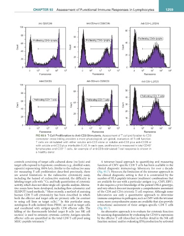
CHaPter 93  Assessment of Functional Immune Responses in Lymphocytes                    1259


                           Anti-CD3/CD45                  Anti-CD3+anti-CD28/CD45              Anti-CD3+IL-2/CD45
              100
                                                                                    60
               80
                                                  60                                         Proliferating CD45+ lymphs
                       Proliferating CD45+ lymphs         Proliferating CD45+ lymphs
               60                                                                   40
             Count                              Count  40                          Count

               40
                                                                                    20
                                                  20
               20

                0                                  0                                 0
                    0    10 0   10 1  10 2  10 3        0   10 0   10 1  10 2  10 3       0    10 0  10 1  10 2  10 3
                           Fluorescence                       Fluorescence                       Fluorescence

                           Anti-CD3/CD3                    Anti-CD3+anti-CD28/CD3              Anti-CD3+IL-2/CD3
              100
                                                  60                                50
               80
                                                                                               Proliferating CD3+ T cells
                                                                                    40
                        Proliferating CD3+ T cells         Proliferating CD3+ T cells
               60                                 40
             Count                               Count                             Count  30
               40
                                                                                    20
                                                  20
               20
                                                                                    10
                0                                  0                                 0
                    0    10 0   10 1  10 2  10 3       0    10 0   10 1  10 2  10 3       0    10 0  10 1  10 2  10 3
                           Fluorescence                       Fluorescence                       Fluorescence
                         FIG 93.5  T-Cell Proliferation to Anti-CD3 Stimulants. Assessment of T-cell proliferation to CD3
                         coreceptor cross-linking provides a more physiological, yet global, evaluation of T-cell function.
                         T cells are stimulated with either soluble anti-CD3 alone or soluble anti-CD3 plus anti-CD28 or
                                                                                                     +
                         with soluble anti-CD3 plus interleukin (IL)-2. In each case, proliferation is measured in total CD45
                         lymphocytes and CD3  T cells. An example of anti-CD3–stimulated T-cell response is shown in
                                           +
                         a healthy donor.
           controls consisting of target cells cultured alone (no lysis) and   A tetramer-based approach to quantifying and measuring
           target cells exposed to hypotonic conditions (e.g., distilled water,   function of CMV-specific CD8 T cells has been available in the
           saponin) representing 100% lysis. Similar to the radioactive assay   clinical diagnostic immunology laboratory for over a decade
           for measuring T-cell proliferation described previously, there   (Fig. 93.7). However, the limitation of the tetramer approach in
           are several limitations to the radioactive cytotoxicity assay,   the clinical diagnostic setting is that it is constrained by the
           including the hazard of radioactive material, the difficulty in   number of HLA-peptide tetramer (multimer) combinations that
                              51
           labeling target cells with  Cr, and bulk quantitation of cytotoxic   are available for use with a particular antigen (e.g., CMV, EBV).
           activity, which does not allow single cell–specific analysis. Alterna-  It also requires a priori knowledge of the patient’s HLA genotype,
           tive assays have been developed, including flow cytometry and   and very often it does not incorporate a comprehensive assessment
                               12
           ELISPOT-based methods.  More recently, a method of assessing   of the CD8 and CD4 cytotoxic T-cell response. Although some
           human  CD8  T-cell  cytotoxicity  has  been  described  in which   laboratories use only a quantitative approach to determine
           both the effector and target cells are primary cells (in contrast   immune competence to pathogens, such as CMV with the tetramer
                                       13
           to  using  cell lines  as  target cells).   In this  particular  assay,   assay, more comprehensive assays are available that also provide
           autologous B cells isolated from PBMC are used as target cells   a functional assessment of these antigen-specific CD8 T cells
           and  cocultured  with  antigen-specific  CD8  effector  cells.  The   (Fig. 93.7).
           killing of the fluorescently labeled target B cells (see NK cell   An alternative approach is to evaluate for cytotoxic function
           section) is used to estimate cytotoxic activity. Antigen-specific   by assessing degranulation by evaluating for CD107a expression
           effector cells are quantified in the total CD8 T-cell pool using   by the effector T cell (described in further detail in the NK cell
           MHC-peptide tetramers. 13                              function section) and/or evaluating IFNγ production by activated