
Page 1303 - Clinical Immunology_ Principles and Practice ( PDFDrive )
P. 1303
CHaPter 93 Assessment of Functional Immune Responses in Lymphocytes 1265
major subpopulations of Treg cells include natural Treg (nTreg)
cells that are produced in the thymus and induced Treg (iTreg)
cells that are generated in the periphery from conventional
+
30
-
FOXP3 CD4 T cells. The dysfunction of Treg cells results in
severe autoimmunity, with IPEX serving as the classic prototype
3,31
(Chapter 35). Besides their role in controlling development of
autoimmunity, lack of Treg cells or abnormal Treg function has
been implicated in the etiopathogenesis of graft-versus-host
disease (GVHD) and allograft rejection, while its presence and
32
normal function have been shown to promote allograft tolerance
in solid-organ transplantation. 33,34
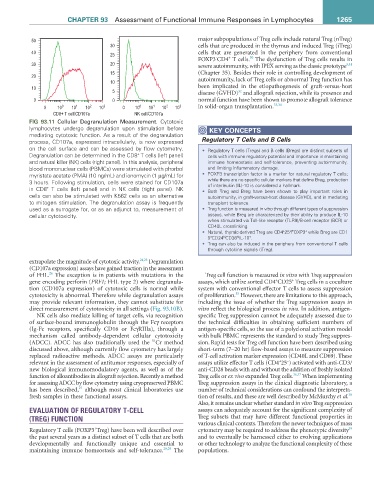
&' 7 FHOO &' D 1. FHOO &' D
FIG 93.11 Cellular Degranulation Measurement. Cytotoxic
lymphocytes undergo degranulation upon stimulation before KeY COnCePtS
mediating cytotoxic function. As a result of the degranulation
process, CD107a, expressed intracellularly, is now expressed Regulatory T Cells and B Cells
on the cell surface and can be assessed by flow cytometry. • Regulatory T cells (Tregs) and B cells (Bregs) are distinct subsets of
+
Degranulation can be determined in the CD8 T cells (left panel) cells with immune regulatory potential and importance in maintaining
and natural killer (NK) cells (right panel). In this analysis, peripheral immune homeostasis and self-tolerance, preventing autoimmunity,
blood mononuclear cells (PBMCs) were stimulated with phorbol and limiting inflammatory damage.
myristate acetate (PMA) (10 ng/mL) and ionomycin (1 µg/mL) for • FOXP3 transcription factor is a marker for natural regulatory T cells;
3 hours. Following stimulation, cells were stained for CD107a while there are no specific cellular markers that define Breg, production
of interleukin (IL)-10 is considered a hallmark.
+
in CD8 T cells (left panel) and in NK cells (right panel). NK • Both Treg and Breg have been shown to play important roles in
cells can also be stimulated with K562 cells as an alternative autoimmunity, in graft-versus-host disease (GVHD), and in mediating
to mitogen stimulation. The degranulation assay is frequently transplant tolerance.
used as a surrogate for, or as an adjunct to, measurement of • Treg function is measured in vitro through different types of suppression
cellular cytotoxicity. assays, while Breg are characterized by their ability to produce IL-10
when stimulated via Toll-like receptor (TLR9)/B-cell receptor (BCR) or
CD40L crosslinking.
+
+
+
• Natural, thymic-derived Treg are CD4 25 FOXP3 while Breg are CD1
hi
hi
+
9 CD24 CD38 IL-10 . +
• Treg can also be induced in the periphery from conventional T cells
through cytokine signals (iTreg).
extrapolate the magnitude of cytotoxic activity. 24,25 Degranulation
(CD107a expression) assays have gained traction in the assessment
26
of FHL. The exception is in patients with mutations in the Treg cell function is measured in vitro with Treg suppression
+
+
gene encoding perforin (PRF1; FHL type 2) where degranula- assays, which utilize sorted CD4 CD25 Treg cells in a coculture
tion (CD107a expression) of cytotoxic cells is normal while system with conventional effector T cells to assess suppression
35
cytotoxicity is abnormal. Therefore while degranulation assays of proliferation. However, there are limitations to this approach,
may provide relevant information, they cannot substitute for including the issue of whether the Treg suppression assays in
direct measurement of cytotoxicity in all settings (Fig. 93.10B). vitro reflect the biological process in vivo. In addition, antigen-
NK cells also mediate killing of target cells, via recognition specific Treg suppression cannot be adequately assessed due to
of surface-bound immunoglobulin through the Fcγ receptors the technical difficulties in obtaining sufficient numbers of
(Ig-Fc receptors, specifically CD16 or FcγRIIIa), through a antigen-specific cells, so the use of a polyclonal activation model
mechanism called antibody-dependent cellular cytotoxicity with bulk PBMC represents the standard to study Treg suppres-
51
(ADCC). ADCC has also traditionally used the Cr method sion. Rapid tests for Treg cell function have been described using
discussed above, although currently flow cytometry has largely short-term (7–20 hr) flow-based assays to measure suppression
replaced radioactive methods. ADCC assays are particularly of T-cell activation marker expression (CD40L and CD69). These
+
−
relevant in the assessment of antitumor responses, especially of assays utilize effector T cells (CD4 25 ) activated with anti-CD3/
new biological immunomodulatory agents, as well as of the anti-CD28 beads with and without the addition of freshly isolated
function of alloantibodies in allograft rejection. Recently a method Treg cells or ex vivo expanded Treg cells. 36,37 When implementing
for assessing ADCC by flow cytometry using cryopreserved PBMC Treg suppression assays in the clinical diagnostic laboratory, a
27
has been described, although most clinical laboratories use number of technical considerations can confound the interpreta-
38
fresh samples in these functional assays. tion of results, and these are well described by McMurchy et al.
Also, it remains unclear whether standard in vitro Treg suppression
EVALUATION OF REGULATORY T-CELL assays can adequately account for the significant complexity of
(TREG) FUNCTION Treg subsets that may have different functional properties in
various clinical contexts. Therefore the newer techniques of mass
+
39
Regulatory T cells (FOXP3 Treg) have been well described over cytometry may be required to address the phenotypic diversity
the past several years as a distinct subset of T cells that are both and to eventually be harnessed either to evolving applications
developmentally and functionally unique and essential to or other technology to analyze the functional complexity of these
maintaining immune homeostasis and self-tolerance. 28,29 The populations.