
Page 1303 - Hall et al (2015) Principles of Critical Care-McGraw-Hill
P. 1303
910 PART 7: Hematologic and Oncologic Disorders
common complications are neurologic (11% of episodes), including the rate at which it develops determine the need for ICU admission,
seizures or stroke. Cardiac, gastrointestinal, or renal complications are emergency transfusion, and aggressive respiratory support. 28
infrequent. Mean duration of hospitalization was 10.5 days, as compared Transfusion therapy is an important consideration in any case of ACS.
with 3 to 4 days for uncomplicated ACS. The outcomes for patients Simple transfusion of 2 to 4 U of packed red blood cells should be con-
17
with ACS may be improving in the current era of aggressive and early sidered early when ACS is diagnosed. Simple transfusion appears to be
transfusion therapy. In the recently completed DeNOVO clinical trial as effective as a more complete red blood cell exchange, but hemoglobin
of inhaled NO gas for patients with acute VOC, only 10% of patients must be maintained below 10 g/dL. This is usually possible because
29
admitted in VOC went on to develop ACS. All patients with ACS were hemoglobin levels usually decrease during ACS. In the event of rapid
24
transfused and transferred to medical intensive care units and none progression of respiratory distress, more severe hypoxemia, multilobar
required mechanical ventilation or died. disease, or requirement for mechanical ventilation, exchange transfu-
Treatment of ACS involves careful supportive care, empiric antibiotic sion is more definitive, more rapid, and the current standard of care (see
therapy, and transfusion therapy (Table 96-8). Pain should be treated section on transfusion therapy). Simple transfusion can be performed as
aggressively, and often patient-controlled analgesia is best. Relief of chest a temporizing maneuver until manual or automated exchange transfu-
pain may improve tidal volume and improve oxygenation, although it sion can be performed.
is important to avoid excessive sedation and respiratory suppression. Other treatments for ACS have been reported in the literature.
Incentive spirometry also helps to reduce atelectasis. Because an infec- Dexamethasone pulse therapy (0.3 mg/kg intravenously every 12 hours in
tious etiology in practice can rarely be ruled out, initial management four doses) has been reported to be effective in decreasing the severity of
should include empiric antibiotic coverage for Chlamydia pneumoniae, ACS in children. However, the rate of rebound pain or ACS progression
Mycoplasma pneumoniae, and Streptococcus pneumoniae, frequently when dexamethasone is stopped may exceed 25%, thus preventing wide-
encountered organisms in ACS. Commonly a third-generation cepha- spread acceptance of this treatment. Our own anecdotal experience indi-
30
17
losporin is combined with a macrolide or quinolone antibiotic. Patients cates that a steroid taper does not alleviate this rebound. Dexamethasone
may respond to inhaled bronchodilator therapy, especially if they have a may be useful in clinical settings in which transfusion therapy is not avail-
history of reactive airway disease. 16 able or as a temporizing measure pending definitive therapy with exchange
Pulse oximetry is a useful tool to monitor the severity of hypoxemia. transfusion. Two case series have suggested that inhaled NO might provide
Due to difficulties in interpretation caused by sickle cell disease, the data clinical benefit, but this cannot be recommended without further clinical
from pulse oximetry are only approximate. 25-27 Pulse oximetry while the investigation in patients with sickle cell disease. 31,32 A recently completed
patient is on room air should be followed, with consideration of arterial randomized-placebo controlled trial of inhaled NO for patients in VOC
blood gas sampling while the patient is on room air if the oxygen satura- found no benefit for this therapy. There was no reduction in the duration of
tion is below 92%. An increase in the alveolar-arterial oxygen gradient hospitalization, the severity of pain, or in the number of patients who went
is the best predictor of ACS severity. The magnitude of the gradient and on to develop ACS during the hospitalization. 24
■ PULMONARY HYPERTENSION
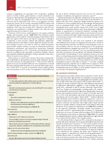
TABLE 96-8 Therapy of the Acute Chest Syndrome of Sickle Cell Disease This frequent but previously underreported complication of sickle cell dis-
ease is associated with a high mortality rate. Population screening studies in
Judicious hydration
adults using noninvasive Doppler-echocardiography have reported a preva-
1-1.5 times daily requirement; fluid restriction may be indicated in patients with severe lence as high as 30% with an associated 10-fold increased risk for death. A
33
acute chest syndrome and capillary leak or in renal insufficiency retrospective analysis of patients diagnosed with pulmonary hypertension
Oxygen by right heart catheterization suggested a 2-year mortality rate appro-
aching 50%, comparable to that of primary pulmonary hypertension.
15
Indicated to maintain adequate oxygenation; does not offer benefit for vaso-occlusive
crisis in the absence of hypoxemia Three new Doppler-echocardiographic screening studies with confirma-
tion of pulmonary hypertension by right heart catheterization (mean pul-
Pain management monary artery pressures of greater than or equal to 25 mm Hg) report that
Mild pain 6% to 10.5% of all adult patients with sickle cell disease have pulmonary
Oral codeine, acetaminophen or ibuprofen hypertension, and are at the highest risk for death. 34-37 Sudden death from
sickle cell disease may be linked to pulmonary hypertension. Severity of
Moderate to severe pain
pulmonary hypertension in patients with sickle cell disease in steady state
Medication can be administered on a fixed time schedule with interval analgesics to is mild to moderate and associated with a high cardiac output and low
obtain adequate pain control (see Table 96–9) pulmonary vascular resistance compared with control subjects with sickle
Consider patient controlled analgesia (see Table 96–10) cell disease without pulmonary hypertension (see Tables 96-3 and 96-4).
In our experience, pulmonary vasculature remains vaso-responsive to
Prevent atelectasis
prostacyclin and NO in approximately 80% of patients. CT angiographic
Incentive spirometry: 10 maximum inspirations q2h while awake images from an 18-year-old patient with homozygous Hb-SS disease with
Empiric antibiotics severe pulmonary hypertension are illustrated in Figure 96-6. The patient
has enlarged pulmonary arteries, a dilated right atrium and right ventricle,
Cephalosporin to cover Streptococcus pneumoniae
and a mosaic perfusion pattern in the lung parenchyma.
Include macrolide or quinolone for coverage of atypical pathogens Chlamydia Our data indicated that endogenous NO is consumed in the plasma of
pneumoniae and Mycoplasma pneumoniae patients with sickle cell disease by the cell-free hemoglobin liberated from
Cultures should include nasal washings for viral pathogens (influenza, respiratory red cells by intravascular hemolysis. 8,38-40 The increase in plasma hemo-
syncytial virus, adenovirus, parainfluenza virus, cytomegalovirus, and parvovirus) globin commonly observed during VOC may heighten NO consumption
during crisis and cause acute decompensation of previously moderate
Diagnose and treat reactive airways disease
pulmonary hypertension. This is exacerbated by an apparent depletion
Consider occult positive end-expiration pressure and its complications of the substrate for NO synthesis, the amino acid l-arginine, by the
Consider exchange transfusion or simple transfusion (see Table 96–12) enzyme arginase-1, released by the red blood cell during hemolysis. 38,39
Inhaled nitric oxide Elevated plasma levels of endothelin 1, an extremely potent vasocon-
strictor, are found in patients with sickle cell disease during VOC, poten-
May prove efficacious but cannot be recommended routinely at this time
tially increasing pulmonary artery pressures in VOC. Chronic and acute
section07.indd 910 1/21/2015 7:43:25 AM