
Page 1301 - Hall et al (2015) Principles of Critical Care-McGraw-Hill
P. 1301
908 PART 7: Hematologic and Oncologic Disorders
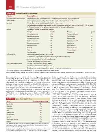
TABLE 96-6 Pathogenesis of the Acute Chest Syndrome
Mechanism Supporting Evidence
Bone infarction leads to atelectasis and Rib, vertebral, and sternal bone infarctions result in pain, hypoventilation, atelectasis, and subsequent hypoxia
regional hypoxia Incentive spirometry decreases radiographic atelectasis in patients with sickle cell anemia and VOC
Fat emboli Evidence of bone marrow embolization found in 9%-75% of autopsy series
Lipid-laden alveolar macrophages can be recovered from 20%-60% of patients with ACS; sPLA levels are elevated in ACS; sPLA may liberate
2
2
free fatty acids from bone marrow lipid, releasing arachidonic acid and promulgating inflammation
Infection Microbiological, serologic, or PCR evidence of pathogens a
Pathogen Episodes Pathogen Episodes
Chlamydia pneumoniae 29% Haemophilus influenzae 2%
Mycoplasma pneumoniae 20% Cytomegalovirus 2%
Respiratory syncytial virus 10% Influenza A virus 2%
Staphylococcus aureus 5% Legionella pneumophila 2%
Streptococcus pneumoniae 4% Escherichia coli 1%
Mycoplasma hominis 4% Epstein-Barr virus 1%
Parvovirus 4% Herpes simplex virus 1%
Rhinovirus 3% Pseudomonas species 1%
Parainfluenza virus 2% Miscellaneous 6%
Vascular occlusion Increased adherence of erythrocytes to endothelial cells
In animal models, regional pulmonary hypoxia results in entrapment of sickle erythrocytes
Vascular obstruction indicated by ventilation/perfusion scan
Pulmonary emboli documented by autopsy series
Vascular injury and inflammation Endothelin 1 levels are elevated during VOC and ACS
Elevated levels of inflammatory mediators such as sPLA
2
Clinical progression to adult respiratory distress syndrome (noncardiogenic pulmonary edema)
a ACS, acute chest syndrome; sPLA ; secretory phospholipase A ; PCR, polymerase chain reaction; VOC, vaso-occlusive pain crisis.
2
2
Data from Vichinsky EP, Neumayr LD, Earles AN, et al. Causes and outcomes of the acute chest syndrome in sickle cell disease. National Acute Chest Syndrome Study Group. N Engl J Med. June 22, 2000;342(25):1855-1865.
those classically seen in patients with femur and pelvic fractures after This receptor, normally involved in the recruitment of inflammatory
trauma (Figs. 96-4 and 96-5). Serum levels of secretory phospholipase cells, may bind erythrocytes and leukocytes, contributing to pulmonary
A rise before the clinical onset of ACS associated with painful crisis. vaso-occlusion. This pulmonary microvascular obstruction worsens
19
2
This enzyme can hydrolyze phospholipids into potent inflammatory ventilation- perfusion mismatch, thereby aggravating hypoxemia, which
mediators such as free fatty acids and lysophospholipids. Liberation of increases sickling and leads to a vicious cycle (see Fig. 96-5). The ACS
arachidonic acid may lead to production of leukotrienes, thrombox- can evolve to a common end point of acute lung injury resembling that
anes, and prostaglandins, all of which mediate inflammation and affect of ARDS, regardless of the initial etiology. 16,22 Interestingly, a recent
endothelial function. Inflammation leads to endothelial cell surface study found that approximately 17% of patients with ACS had an abnor-
expression of cell adhesion molecules, especially VCAM-1. Expression mal CT-angiogram of the pulmonary vasculature, suggesting acute
of VCAM-1 may be further increased by depletion of NO and its thromboembolism or in situ thrombosis. 23
precursor arginine, which normally suppress VCAM-1 expression. 20,21 The clinical severity of ACS is highly variable. Common physical
findings include fever, tachypnea, rales, and wheezing. Laboratory
findings often include leukocytosis, an acute decrease in hemoglobin
TABLE 96-7 Causes of Acute Chest Syndrome a
level (average decrease of approximately 1.6 g/dL) and platelet count
Cause Cases With Identified Etiology (often dropping from a baseline of about 400,000/mm to less than
3
3
Fat embolism 16% 200,000/mm ), and hypoxemia. Pulmonary infiltrates are often found in
Chlamydia 13% the upper lobes in children and lower lobes in adults. Multilobar disease
suggests a worse prognosis. In addition to a new pulmonary infiltrate
Mycoplasma 12% or consolidation, pleural effusions develop in up to half of the episodes
Virus 12% during the course of hospitalization (see Fig. 96-4). In the study by the
Typical bacteria 8% National Acute Chest Syndrome Study Group, 22% of ACS episodes
Mixed infections 7% in adults and 10% of episodes in children required management with
Legionella 1% mechanical ventilation. Risk factors for requiring mechanical ventila-
tion were decreased platelet count (<200,000/mm ), multilobar disease,
3
Miscellaneous infections 1% a history of cardiac problems, and neurologic complications. Of sickle
Infarction 30% cell patients admitted to the ICU for ACS, 60% develop abnormally high
a Only those cases with complete analysis for the indicated potential infectious causes by culture, serol- pulmonary artery pressures estimated by echocardiography, and those
ogy, and polymerase chain reaction are reported. Cases without any defined infectious etiology were with tricuspid regurgitant velocity above 3 m/s and evident cor pulmo-
presumed to be due to infarction without infection. nale (right heart failure) were most likely to develop respiratory failure
Data from Vichinsky EP, Neumayr LD, Earles AN, et al. Causes and outcomes of the acute chest syndrome in sickle and die. Total mortality rates were 9% in adults but lower than 1% in
cell disease. National Acute Chest Syndrome Study Group. N Engl J Med. June 22, 2000;342(25):1855-1865. children. Other than pain (especially abdominal or chest pain), the most
section07.indd 908 1/21/2015 7:43:22 AM