
Page 1375 - Hall et al (2015) Principles of Critical Care-McGraw-Hill
P. 1375
948 PART 8: Renal and Metabolic Disorders
3. Elevated urine sodium
TABLE 99-3 Etiologies of Hyponatremia Categorized by Clinical Volume Status
4. No underlying adrenal, thyroid, pituitary, or renal disease
Hypovolemic Euvolemic Hypervolemic
Diarrhea SIADH CHF Unique Clinical Situations Resulting in Hyponatremia
Vomiting Hypothyroidism Cirrhosis Cerebral Salt Wasting Cerebral salt wasting (CSW) is a rare clinical event that
typically follows a subarachnoid hemorrhage. Patients have high urinary
Pancreatitis Glucocorticoid deficiency Nephrotic syndrome
flow rates with elevated urine sodium, which ultimately results in severe
Burns Acute renal failure volume deficiency and hyponatremia. The pathophysiology behind CSW
Diuretic induced Chronic renal failure has not been fully elucidated but it is likely that natriuretic proteins are
released in response to the CNS injury. Atrial natriuretic peptide, brain
Mineralocorticoid deficiency
natriuretic peptide, and an endogenous ouabain-like peptide have all
Salt-wasting nephropathy been proposed as possible etiologic agents. The key diagnostic dilemma
Cerebral salt wasting is differentiating CSW from SIADH, as both can follow CNS insults and
are marked by hyponatremia with elevated urine sodium. The principal
CHF, congestive heart failure; SIADH, the syndrome of inappropriate secretion of antidiuretic hormone.
differences are urine volume (in SIADH it is low, while it is high in CSW)
and clinical volume status (it is normal in SIADH, while it is low in
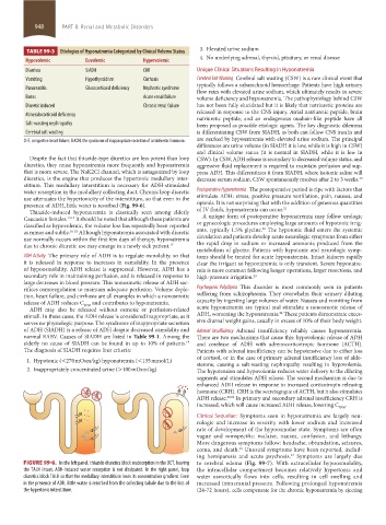
Despite the fact that thiazide-type diuretics are less potent than loop CSW). In CSW, ADH release is secondary to decreased volume status, and
diuretics, they cause hyponatremia more frequently and hyponatremia aggressive fluid replacement is required to maintain perfusion and sup-
that is more severe. The NaK2Cl channel, which is antagonized by loop press ADH. This differentiates it from SIADH, where isotonic saline will
diuretics, is the engine that produces the hypertonic medullary inter- decrease serum sodium. CSW spontaneously resolves after 2 to 3 weeks. 20
stitium. This medullary interstitium is necessary for ADH-stimulated
water resorption in the medullary collecting duct. Chronic loop diuretic Postoperative Hyponatremia The postoperative period is ripe with factors that
use attenuates the hypertonicity of the interstitium, so that even in the stimulate ADH: stress, positive pressure ventilation, pain, nausea, and
presence of ADH, little water is resorbed (Fig. 99-6). opioids. It is not surprising that with the addition of generous quantities
21
Thiazide-induced hyponatremia is classically seen among elderly of IV fluids, hyponatremia can occur.
Caucasian females. 14,15 It should be noted that although these patients are A unique form of postoperative hyponatremia may follow urologic
classified as hypovolemic, the volume loss has repeatedly been reported or gynecologic procedures employing large amounts of hypotonic irrig-
22
as minor and subtle. 16-18 Although hyponatremia associated with diuretic ants, typically 1.5% glycine. The hypotonic fluid enters the systemic
use normally occurs within the first few days of therapy, hyponatremia circulation and patients develop acute neurologic symptoms from either
due to chronic diuretic use may emerge in a newly sick patient. 17 the rapid drop in sodium or increased ammonia produced from the
metabolism of glycine. Patients with hypotonic and neurologic symp-
ADH Activity The primary role of ADH is to regulate osmolality, so that toms should be treated for acute hyponatremia. Intact kidneys rapidly
it is released in response to increases in osmolality. In the presence clear the irrigant so hyponatremia is only transient. Severe hyponatre-
of hypoosmolality, ADH release is suppressed. However, ADH has a mia is more common following longer operations, larger resections, and
secondary role in maintaining perfusion, and is released in response to high-pressure irrigation. 23
large decreases in blood pressure. This nonosmotic release of ADH sac-
rifices osmoregulation to maintain adequate perfusion. Volume deple- Psychogenic Polydipsia This disorder is most commonly seen in patients
tion, heart failure, and cirrhosis are all examples in which a nonosmotic suffering from schizophrenia. They overwhelm their urinary diluting
release of ADH reduces C EFW and contributes to hyponatremia. capacity by ingesting large volumes of water. Nausea and vomiting from
ADH may also be released without osmotic or perfusion-related acute hyponatremia are typical and stimulate a nonosmotic release of
24
stimuli. In these cases, the ADH release is considered inappropriate, as it ADH, worsening the hyponatremia. These patients demonstrate exces-
serves no physiologic purpose. The syndrome of inappropriate secretion sive diurnal weight gains, usually in excess of 10% of their body weight.
of ADH (SIADH) is a release of ADH despite decreased osmolality and Adrenal Insufficiency Adrenal insufficiency reliably causes hyponatremia.
normal EABV. Causes of SIADH are listed in Table 99-1. Among the There are two mechanisms that cause this: hypovolemic release of ADH
19
elderly no cause of SIADH can be found in up to 10% of patients. and corelease of ADH with adrenocorticotropic hormone (ACTH).
The diagnosis of SIADH requires four criteria: Patients with adrenal insufficiency can be hypotensive due to either loss
of cortisol, or in the case of primary adrenal insufficiency loss of aldo-
1. Hypotonic (<270 mOsm/kg) hyponatremia (<135 mmol/L) sterone, causing a salt-wasting nephropathy resulting in hypovolemia.
2. Inappropriately concentrated urine (>100 mOsm/kg) The hypotension and hypovolemia reduces water delivery to the diluting
segments and stimulates ADH release. The second mechanism is due to
enhanced ADH release in response to increased corticotropin-releasing
hormone (CRH). CRH is the secretagogue of ACTH, but it also stimulates
ADH release. 25,26 In primary and secondary adrenal insufficiency CRH is
increased, which will cause increased ADH release, lowering C .
EFW
Clinical Sequelae: Symptoms seen in hyponatremia are largely neu-
rologic and increase in severity with lower sodium and increased
rate of development of the hypoosmolar state. Symptoms are often
vague and nonspecific: malaise, nausea, confusion, and lethargy.
More dangerous symptoms follow: headache, obtundation, seizures,
coma, and death. Unusual symptoms have been reported, includ-
16
ing hemiparesis and acute psychosis. Symptoms are largely due
27
FIGURE 99-6. In the left panel, thiazide diuretics block reabsorption in the DCT, leaving to cerebral edema (Fig. 99-7). With extracellular hypoosmolality,
the TALH intact. ADH-induced water resorption is not dissipated. In the right panel, loop the intracellular compartment becomes relatively hypertonic and
diuretics block TALH so that the medullary interstitium loses its concentration gradient. Even water osmotically flows into cells, resulting in cell swelling and
in the presence of ADH, little water is resorbed from the collecting tubule due to the loss of increased intracranial pressure. Following prolonged hyponatremia
the hypertonic interstitium. (24-72 hours), cells compensate for the chronic hyponatremia by ejecting
section08.indd 948 1/14/2015 8:28:11 AM