
Page 1379 - Hall et al (2015) Principles of Critical Care-McGraw-Hill
P. 1379
952 PART 8: Renal and Metabolic Disorders
diuresis in response to the hypertension known as aldosterone escape. A inappropriately wastes potassium. Three studies may be used to differ-
full discussion of the causes of increased aldosterone activity is beyond entiate these states: spot urine potassium concentration, 24-hour urine
the scope of this text; however, a list of causes is included in Table 99-4. potassium, and the transtubular potassium gradient (TTKG).
Normally, the primary anion in the tubular fluid is chloride. Various The spot urine is the simplest test to use. The urine potassium should
conditions can result in chloride being replaced by an unresorb- be less than 20 mmol/L in the face of hypokalemia. If the spot potas-
able anion. Anions that are not resorbed prevent sodium from being sium is greater than 40, renal potassium wasting should be suspected.
resorbed and increase sodium and tubular fluid delivery to the distal Urine potassium of 20 to 40 mmol/L is considered nondiagnostic.
81
nephron. In addition, unresorbable anions increase tubule electronega- There are two primary problems with this test; the first is it fails to
tivity, which enhances potassium secretion by the principal cells. The control for changes in the water content of urine. Since hypokalemia
most common example of an unresorbable anion resulting in hypokale- is associated with decreased ADH sensitivity, increased water content
mia is bicarbonate. In metabolic alkalosis, increased serum bicarbonate will lower the urinary potassium concentration. The second problem
is delivered to the distal nephron, resulting in increased renal potassium is that spot samples provide information for only a single moment
loss. Diabetic ketoacidosis increases delivery of the unresorbable anion in time. Patients with diuretic-induced hypokalemia become potas-
β-hydroxybutyrate to the distal nephron. sium avid after the diuretic has cleared. One study on the diagnosis
Hypomagnesemia is associated with hypokalemia that is resistant to of hypokalemia (mean K = 2.0 mmol/L) found spot urine potassium
therapy. Decreased magnesium increases renal potassium losses and to have a sensitivity of 40% and specificity of 100% for excess renal
needs to be corrected prior to successful treatment of hypokalemia. 70 potassium loss. 82
Despite a high concentration of potassium in lower GI secretions, The 24-hour urine potassium test avoids both of the above prob-
85 to 95 mmol/L, GI potassium losses are typically modest, about lems at the expense of increased complexity and a 24-hour delay.
10 mEq/d. Chronic diarrhea can cause hypokalemia, but the mecha- Patients with hypokalemia should reduce urinary potassium losses
51
nism appears to be more complex than simple GI loss of potassium. In to less than 15 mEq/d. Potassium losses greater than that indicate
cases of experimental diarrhea, daily GI potassium loss was never higher inappropriate renal losses. The 24-hour urine provides no informa-
than 24 mEq/d, a level well below average daily potassium intake. In tion on the renal potassium handling prior to the urine collection
71
addition, studies on diarrhea show that as stool volume increases, stool (eg, diuretic use that is stopped prior to collection will show an
potassium concentration falls, ultimately reaching a level similar to that appropriately potassium-avid kidney).
of plasma in cases of severe cholera. Explanations for the commonly The transtubular potassium gradient calculates the ratio of tubular
49
seen association of diarrhea and hypokalemia include secondary hyper- potassium to venous potassium at the end of the CCD. The CCD is
aldosteronism, diminished intake of potassium, or transcellular shifts of responsible for potassium excretion, so increases in the TTKG indicate
extracellular potassium. renal wasting of potassium, while decreases indicate renal potassium
Gastric secretions have potassium content similar to that of plasma, 5 conservation (Fig. 99-10 and Eq. 99-5). When serum and renal potas-
to 8 mmol/L. Gastric losses result in severe metabolic alkalosis and sec- sium handling are normal, the TTKG runs from 5 to 8. 81,83 In the face
ondary hyperaldosteronism, both of which enhance renal potassium loss. of hypokalemia, the CCD should minimize the potassium excretion,
resulting in a reduced TTKG. The TTKG has been validated in patients
Clinical Sequelae: Hypokalemia is a well-known risk factor for a variety with decreased dietary potassium, periodic paralysis, diuretic-induced
of cardiac arrhythmias. Increased ectopy with hypokalemia has been hypokalemia, primary hyperaldosteronism, and vomiting. 83,84
documented in ambulatory hypertensive patients, in patients under-
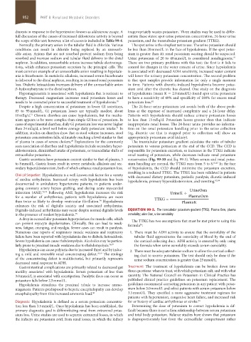
going coronary artery bypass grafting, and during acute myocardial UrineOsm
infarction (AMI). 72,73 Following AMI, hypokalemia increases the risk UrineK ÷
for a number of arrhythmias; patients with hypokalemia are more TTKG= PlasmaOsm
than twice as likely to develop ventricular fibrillation. Hypokalemia PlasmaK
74
enhances the risk of digitalis toxicity and associated arrhythmias.
Digitalis-induced arrhythmias may occur despite normal digitalis levels Equation 99-5. The transtubular potassium gradient (TTKG). Plasma Osm, plasma
in the presence of modest hypokalemia. 75 osmolality; urine Osm, urine osmolality.
A drop in extracellular potassium hyperpolarizes the muscle cells, which The TTKG has two assumptions that must be met prior to using this
can prevent myocyte depolarization. Clinically, this can lead to weak- formula :
85
ness, fatigue, cramping, and myalgia. Severe cases can result in paralysis.
Numerous case reports of respiratory muscle weakness and respiratory 1. There must be ADH activity to ensure that the osmolality of the
failure have been reported with hypokalemia due to diabetic ketoacidosis. tubular fluid approximates the osmolality of blood by the end of
Severe hypokalemia can cause rhabdomyolysis. Alcoholics may be particu- the cortical collecting duct. ADH activity is ensured by only using
larly prone to proximal muscle weakness due to rhabdomyolysis. 76,77 the formula when urine osmolality exceeds serum osmolality.
Hypokalemia can cause polyuria due to increased thirst and by induc- 2. There must be adequate tubular sodium to allow the cortical collect-
ing a mild and reversible renal concentrating defect. 78,79 The etiology ing duct to secrete potassium. The test should only be done if the
of the concentrating defect is multifactorial, but primarily represents urine sodium concentration is greater than 25 mmol/L.
decreased renal response to ADH.
Gastrointestinal complications are primarily related to decreased gut Treatment: The treatment of hypokalemia can be broken down into
motility associated with hypokalemia. Serum potassium of less than three questions: when to treat, with which potassium salt, and with what
3.0 mmol/L is associated with constipation. Paralytic ileus can occur as quantity. The National Council on Potassium in Clinical Practice has
potassium falls below 2.5 mmol/L. published clinical practice guidelines on potassium replacement. The
Hypokalemia stimulates the proximal tubule to increase ammo- guidelines recommend correcting potassium in any patient with potas-
niagenesis. Patients predisposed to hepatic encephalopathy can develop sium below 3.0 mmol/L and select patients with serum potassium below
encephalopathy from this increased ammonia load. 80 3.5 mmol/L. They specified a more aggressive treatment regimen for
patients with hypertension, congestive heart failure, and increased risk
Diagnosis: Hypokalemia is defined as a serum potassium concentra- for or history of cardiac arrhythmias or stroke. 86
tion less than 3.5 mmol/L. Once hypokalemia has been established, the Determining the dose of potassium to correct hypokalemia is dif-
primary diagnostic goal is differentiating renal from extrarenal potas- ficult because there is not a firm relationship between serum potassium
sium loss. Urine studies are used to separate extrarenal losses, in which and total body potassium. Balance studies have shown that potassium
the kidneys are potassium avid, from renal losses, in which the kidney is disproportionately lost from the extracellular compartment rather
section08.indd 952 1/19/2015 11:35:07 PM