
Page 1387 - Hall et al (2015) Principles of Critical Care-McGraw-Hill
P. 1387
960 PART 8: Renal and Metabolic Disorders
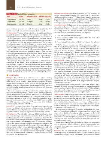
TABLE 99-10 Parenteral Calcium Formulations Endogenous Calcitriol Production Calcitriol synthesis can be increased by
chronic granulomatous disorders (eg, tuberculosis or sarcoidosis),
Agent Supplied Elemental Ca per mL Elemental Ca per Gram lymphomas, and acromegaly. 199,200 Macrophages found in granulomas
Calcium gluconate 1 g in 10 mL 9 mg/mL 90 mg 4.5 mEq convert 25-hydroxyvitamin D to calcitriol despite low PTH levels. Both
Hodgkin disease and non-Hodgkin lymphoma can cause hypercalcemia
Calcium chloride 1 g in 10 mL 27.2 mg/mL 272 mg 13.6 mEq
by endogenous production of 1,25-dihydroxyvitamin D.
Calcium gluceptate 1 g in 5 mL 18 mg/mL 90 mg 4.5 mEq
Increased Bone Resorption Malignancy is the most common cause of inpatient
hypercalcemia. Twenty to thirty percent of patients with cancer develop
access. Calcium gluconate can safely be infused peripherally. Both hypercalcemia. The most common associated malignancies are breast
201
calcium compounds can cause tissue necrosis if extravasated. cancer, lung cancer, and multiple myeloma. There are three primary
Other Treatment Issues Treatment of hypocalcemia can precipitate arrhythmias, mechanisms for increased bone resorption in malignancy:
especially in patients on digitalis. Other complications reported from 1. Local osteolysis from bone metastasis
treating hypocalcemia include bradycardia, pancreatitis, and vasospasm. 196
When hypocalcemia is due to hyperphosphatemia, there is concern 2. Tumor secretion of PTH-related peptide (PTH-rP), often called
that providing calcium could accelerate metastatic soft-tissue calcifica- humoral hypercalcemia
tion. The degree to which this occurs is unclear. In the face of hyper- 3. Tumor-induced hydroxylation of 25-hydroxyvitamin D to calcitriol
phosphatemia, calcium should be limited to reversing acute toxicity PTH-rP is the most common cause of hypercalcemia of malignancy.
(tetany, laryngospasm, and arrhythmias) and full correction of hypocal- PTH-rP is a physiologic protein that is normally involved in the syn-
cemia should be delayed until the phosphorus is normalized. 169 thesis and development of cartilage. PTH-rP causes hypercalcemia
Hypomagnesemia can contribute to hypocalcemia so patients should
have a magnesium level checked and repleted if low. There have been by binding to PTH receptors. Though similar in structure to PTH,
177
PTH-rP is not measured by PTH assays and requires a specific blood
reports of magnesium-responsive hypocalcemia despite normal serum test. Hypercalcemia from PTH-rP is most common in nonmetastatic
magnesium levels. This is thought to be due to total body magnesium solid tumors, non-Hodgkin lymphoma, chronic myeloid leukemia (blast
depletion despite normal serum levels (see section on the diagnosis of phase), and adult T-cell lymphoma.
hypomagnesemia below for details).
The principal therapy for hypocalcemia due to citrate toxicity is Hyperparathyroidism Primary hyperparathyroidism is the most frequent
metabolism of the citrate. Citrate metabolism occurs via tempera- cause of hypercalcemia. Mild hypercalcemia, hypophosphatemia, and
ture-dependent enzymes so correcting hypothermia improves hepatic elevated PTH are the hallmarks of this condition. Generally patients
metabolism. Steps to improve hypotension and hepatic blood flow should have three normal parathyroid glands with one large gland containing a
be taken. Saline loading in order to increase renal clearance may speed functional adenoma. However, in 15% of cases there will be hyperplasia
recovery. Be aware that saline loading will also increase renal calcium of all four glands. Parathyroid cancer accounts for less than 1% of pri-
excretion. About 20% of citrate is excreted unmetabolized in the urine. mary hyperparathyroidism. Surgery to remove the autonomous gland is
■ HYPERCALCEMIA the preferred treatment, and in cases of diffuse four-gland enlargement,
three and a half glands are removed. Rarely, extreme symptomatic
Etiologies: Hypercalcemia is a relatively common clinical finding. hypercalcemia can occur with hyperparathyroidism. This is called
parathyroid crisis and is characterized by mental status changes, severe
Hypercalcemia occurs when calcium enters the vascular compartment
faster than it can be excreted. There are two mechanisms by which hypercalcemia, and very high PTH levels. Surgical removal of the para-
thyroid tissue is indicated.
calcium enters the vascular space: calcitriol-mediated gut absorption
Tertiary hyperparathyroidism occurs in chronic renal failure in which
and bone resorption. Likewise, there are two means by which calcium is chronic PTH stimuli (decreased serum calcium or decreased calcitriol)
removed from the vascular space: deposition in bone or soft tissue and
excretion in urine. result in parathyroid glands that autonomously secrete PTH, resulting in
hypercalcemia. These patients typically have four-gland hyperplasia and
The most common cause of hypercalcemia is primary hyperpara-
thyroidism, while malignancy is a distant second. Among hospitalized are resistant to medical management and require surgery.
patients, however, this ratio is reversed, with cancer accounting for 65% Clinical Sequelae: Mild hypercalcemia is associated with relatively mild,
of cases and hyperparathyroidism 25%. One series found milk-alkali nonspecific symptoms. Patients with primary hyperparathyroidism
syndrome to account for up to 12% of patients hospitalized for hyper- are generally asymptomatic, but may complain of weakness, fatigue,
calcemia, while more recently a prevalence rate of 8.8% was reported for anorexia, depression, vague abdominal pain, and constipation.
non-ESRD hypercalcemic inpatients. 197,198 A summary of the etiologies Gastrointestinal side effects become more severe at higher calcium
of hypercalcemia is listed in Table 99-11. levels. Hypercalcemia has been associated with increased gastrin
Increased Intake Increased dietary intake alone rarely causes hypercalcemia secretion and may predispose patients to peptic ulcers. Severe hyper-
because the kidney is able to increase calcium excretion dramatically. calcemia can cause pancreatitis.
Increased intake causes hypercalcemia in patients with renal failure or Hypercalcemia can cause multiple forms of renal dysfunction. Long-
in patients in whom the kidney is prevented from excreting calcium. standing hypercalcemia predisposes patients to nephrolithiasis. It also
Milk-Alkali Syndrome The milk-alkali syndrome (MAS) is defined by three causes volume depletion by reducing sodium resorption in the TALH
and decreasing the renal response to ADH. Hypercalcemia can cause
concurrent findings, hypercalcemia, metabolic alkalosis, and renal acute renal failure by causing volume depletion or by vasoconstriction,
insufficiency, and is due to the ingestion of calcium and alkali. In the reducing renal blood flow. Long-standing hypercalcemia results in irre-
198
modern era, patients are typically women being treated for osteoporosis versible renal insufficiency.
with calcium carbonate, which supplies both the calcium and the alkali. Mental status changes from mild confusion to psychosis or coma can
Historically, MAS was characterized by hyperphosphatemia due to the occur in severe cases of hypercalcemia. It is clinically important to note
high phosphorus content of milk. In modern MAS, the calcium is a that mental status impairment can persist for days following correction
pharmaceutical product without phosphorus and patients tend to be of hypercalcemia. 202
hypophosphatemic, which stimulates calcitriol production, increasing
calcium absorption. The hypercalcemia typically responds to stopping Treatment: The best treatment for hypercalcemia is to correct the
alkali and calcium ingestion. Additionally, saline infusions and loop underlying etiology. In situations in which this is not possible or specific
diuretics are effective treatments. 198 hypocalcemic therapy is needed, the treatment should focus on the three
section08.indd 960 1/14/2015 8:28:19 AM