


Page 168 - Textbook of Pathology, 6th Edition
P. 168
152 iii) tuberculous salpingitis into peritoneal cavity (tuberculous  hours. However, patients having disseminated tuberculosis
           peritonitis);                                       may show negative test due to release of large amount of
           iv) infected sputum into larynx (tuberculous laryngitis);  tuberculoproteins from the endogenous lesions masking the
           v) swallowing of infected sputum (ileocaecal tuberculosis);  hypersensitivity test. A positive test is indicative of cell-
           and                                                 mediated hypersensitivity to tubercular antigens but does
           vi) renal lesions into ureter and down to trigone of bladder.  not distinguish between infection and disease. The test may
                                                               be false positive in atypical mycobacterial infection and false
           HYPERSENSITIVITY AND IMMUNITY IN TUBER-             negative in sarcoidosis, some viral infections, Hodgkin’s
           CULOSIS.  Hypersensitivity or allergy, and immunity or  disease and fulminant tuberculosis.
     SECTION I
           resistance, play a major role in the development of lesions in  Immunisation against tuberculosis. Protective immuni-
           tuberculosis. Tubercle bacilli as such do not produce any  sation against tuberculosis is induced by injection of
           toxins. Tissue changes seen in tuberculosis are not the result  attenuated strains of bovine type of tubercle bacilli, Bacille
           of any exotoxin or endotoxin but are instead the result of  Calmette-Guérin  (BCG). Cell-mediated immunity with
           host response to the organism which is in the form of  consequent delayed hypersensitivity reaction develops with
           development of cell-mediated hypersensitivity (or type IV  healing of the lesion, but the cell-mediated immunity persists,
           hypersensitivity) and immunity. Both these host responses  rendering the host tuberculin-positive and hence immune.
           develop as a consequence of several lipids present in the
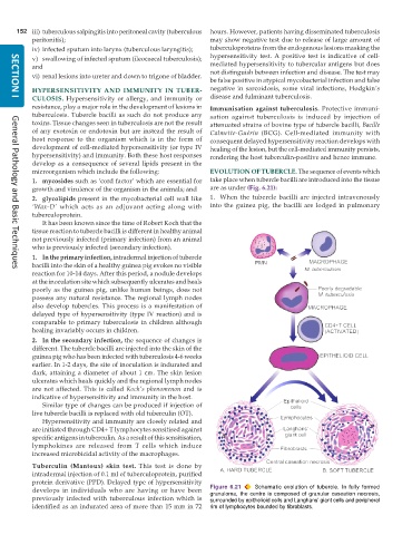
           microorganism which include the following:          EVOLUTION OF TUBERCLE. The sequence of events which
           1. mycosides such as ‘cord factor’ which are essential for  take place when tubercle bacilli are introduced into the tissue
           growth and virulence of the organism in the animals; and  are as under (Fig. 6.21):
           2. glycolipids present in the mycobacterial cell wall like  1. When the tubercle bacilli are injected intravenously
           ‘Wax-D’ which acts as an adjuvant acting along with  into the guinea pig, the bacilli are lodged in pulmonary
           tuberculoprotein.
              It has been known since the time of Robert Koch that the
           tissue reaction to tubercle bacilli is different in healthy animal
           not previously infected (primary infection) from an animal
           who is previously infected (secondary infection).
           1. In the primary infection, intradermal injection of tubercle
           bacilli into the skin of a healthy guinea pig evokes no visible
     General Pathology and Basic Techniques
           reaction for 10-14 days. After this period, a nodule develops
           at the inoculation site which subsequently ulcerates and heals
           poorly as the guinea pig, unlike human beings, does not
           possess any natural resistance. The regional lymph nodes
           also develop tubercles. This process is a manifestation of
           delayed type of hypersensitivity (type IV reaction) and is
           comparable to primary tuberculosis in children although
           healing invariably occurs in children.
           2. In the secondary infection, the sequence of changes is
           different. The tubercle bacilli are injected into the skin of the
           guinea pig who has been infected with tuberculosis 4-6 weeks
           earlier. In 1-2 days, the site of inoculation is indurated and
           dark, attaining a diameter of about 1 cm. The skin lesion
           ulcerates which heals quickly and the regional lymph nodes
           are not affected. This is called  Koch’s phenomenon and is
           indicative of hypersensitivity and immunity in the host.
              Similar type of changes can be produced if injection of
           live tubercle bacilli is replaced with old tuberculin (OT).
              Hypersensitivity and immunity are closely related and
           are initiated through CD4+ T lymphocytes sensitised against
           specific antigens in tuberculin. As a result of this sensitisation,
           lymphokines are released from T cells which induce
           increased microbicidal activity of the macrophages.

           Tuberculin (Mantoux) skin test. This test is done by
           intradermal injection of 0.1 ml of tuberculoprotein, purified
           protein derivative (PPD). Delayed type of hypersensitivity
           develops in individuals who are having or have been  Figure 6.21  Schematic evolution of tubercle. In fully formed
                                                               granuloma, the centre is composed of granular caseation necrosis,
           previously infected with tuberculous infection which is  surrounded by epithelioid cells and Langhans’ giant cells and peripheral
           identified as an indurated area of more than 15 mm in 72  rim of lymphocytes bounded by fibroblasts.

