


Page 166 - Textbook of Pathology, 6th Edition
P. 166
150
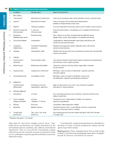
TABLE 6.4: Principal Granulomatous Conditions.
Conditions Etiologic Agent Special Characteristics
I. BACTERIAL
1. Tuberculosis* Mycobacterium tuberculosis Tuberculous granulomas with central caseation necrosis; acid-fast bacilli.
2. Leprosy* Mycobacterium leprae Foamy histiocytes with acid-fast bacilli (lepromatous);
epithelioid cell granulomas (tuberculoid).
3. Syphilis* Treponema pallidum Gummas composed of histiocytes; plasma cell infiltration; central necrosis.
SECTION I
4. Granuloma inguinale C. donovani Anal and genital lesions; macrophages and neutrophils show Donovan
(Donovanosis) (Donovan body) bodies.
5. Brucellosis Brucella abortus Dairy infection to humans; enlarged reticuloendothelial organs
(Mediterranean fever) (lymph nodes, spleen, bone marrow); non-specific granulomas.
6. Cat scratch disease Coccobacillus Lymphadenitis; reticuloendothelial hyperplasia; granulomas with
central necrosis and neutrophils.
7. Tularaemia Francisella (Pasteurella) Necrosis and suppuration (acute); tubercles hard or with minute
(Rabbit fever) tularensis central necrosis (chronic).
8. Glanders Actinobacillus mallei Infection from horses and mules; subcutaneous lesions and lymphadenitis;
infective granulomas.
II. FUNGAL
1. Actinomycosis* Actinomycetes israelii Cervicofacial, abdominal and thoracic lesions; granulomas and abscesses
(bacterial) with draining sinuses; sulphur granules.
2. Blastomycosis Blastomyces dermatitidis Cutaneous, systemic and lung lesions; suppuration; ulceration
and granulomas.
3. Cryptococcosis Cryptococcus neoformans Meninges, lungs and systemic distribution; organism yeast-like
with clear capsule.
General Pathology and Basic Techniques
4. Coccidioidomycosis Coccidioides immitis Meninges, lungs and systemic distribution; granulomas
and abscesses; organism cyst containing endospores.
III. PARASITIC
Schistosomiasis Schistosoma mansoni, Eggs and granulomas in gut, liver, lung; schistosome pigment;
(Bilharziasis) haematobium, japonicum eosinophils in blood and tissue.
IV. MISCELLANEOUS
1. Sarcoidosis* Unknown Non-caseating granulomas (hard tubercles); asteroid and Schaumann
bodies in giant cells.
2. Crohn’s disease Unknown Transmural chronic inflammatory infiltrates; non-caseating
(Regional enteritis) ? Bacteria, ?? Viruses sarcoid-like granulomas.
3. Silicosis Silica dust Lung lesions, fibrocollagenous nodules.
4. Berylliosis Metallic beryllium Sarcoid-like granulomas in lungs; fibrosis; inclusions in giant cells
(asteroids, Schaumann bodies, crystals).
5. Foreign body Talc, suture, oils, wood Non-caseating granulomas with foreign body giant cells; demonstration
granulomas splinter etc. of foreign body.
*Diseases discussed in this chapter.
other than M. tuberculosis complex and M. leprae. Non- Conventionally, atypical mycobacteria are classified on
tuberculous mycobacteria are widely distributed in the the basis of colour of colony produced in culture and the
environment and are, therefore, also called as environmental speed of growth in media:
mycobacteria. They too are acid fast. Occasionally, human Rapid growers. These organisms grow fast on solid media
tuberculosis may be caused by atypical mycobacteria which (within 7 days) but are less pathogenic than others. Examples
are non-pathogenic to guinea pigs and resistant to usual anti-
tubercular drugs. include M. abscessus, M.fortuitum, M. chelonae.

