


Page 169 - Textbook of Pathology, 6th Edition
P. 169
capillaries where an initial response of neutrophils is evoked  on the surface. This is called cold abscess although there are  153
           which are rapidly destroyed by the organisms.       no pus cells in it.
           2. After about 12 hours, there is progressive infiltration by  ii) In tuberculosis of tissues like bones, joints, lymph nodes
           macrophages. This is due to coating of tubercle bacilli with  and epididymis, sinuses are formed and the sinus tracts are
           serum complement factors C2a and C3b which act as   lined by tuberculous granulation tissue.
           opsonins and attract the macrophages. Macrophages   iii) The adjacent granulomas may coalesce together enlarging  CHAPTER 6
           dominate the picture throughout the remaining life of the  the lesion which is surrounded by progressive fibrosis.
           lesions. If the tubercle bacilli are, however, inhaled into the  iv) In the granuloma enclosed by fibrous tissue, calcium salts
           lung alveoli, macrophages predominate the picture from the  may get deposited in the caseous material  (dystrophic
           beginning.                                          calcification) and sometimes the lesion may even get ossified
           3. The macrophages start phagocytosing the tubercle bacilli  over the years.
           and either kill the bacteria or die away themselves. In the
           latter case, they further proliferate locally as well as there is  TYPES OF TUBERCULOSIS
           increased recruitment of macrophages from blood
           monocytes.                                          Lung is the main organ affected in tuberculosis. Depending
                                                               upon the type of tissue response and age, the infection with
           4. As a part of body’s immune response, T and B cells are  tubercle bacilli is of 2 main types:
           activated.  Activated CD4+T cells develop the cell-mediated  A. Primary tuberculosis; and                  Inflammation and Healing
           delayed type hypersensitivity reaction, while B cells result in  B. Secondary tuberculosis.
           formation of antibodies which play no role in body’s defence
           against tubercle bacilli.                           A. Primary Tuberculosis
           5. In 2-3 days, the macrophages undergo structural changes
           as a result of immune mechanisms—the cytoplasm becomes  The infection of an individual who has not been previously
           pale and eosinophilic and their nuclei become elongated and  infected or immunised is called primary tuberculosis or Ghon’s
           vesicular. These modified macrophages resemble epithelial  complex or childhood tuberculosis.
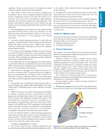
           cells and are called epithelioid cells.                Primary complex or Ghon’s complex is the lesion
           6. The epithelioid cells in time aggregate into tight clusters  produced in the tissue of portal of entry with foci in the
                                                               draining lymphatic vessels and lymph nodes. The most
           or granulomas. Release of cytokines in response to sensitised  commonly involved tissues for primary complex are lungs
           CD4+T cells and some constituents of mycobacterial cell wall  and hilar lymph nodes. Other tissues which may show
           play a role in formation of granuloma.
                                                               primary complex are tonsils and cervical lymph nodes, and
           7. Some of the macrophages form multinucleated giant cells  in the case of ingested bacilli the lesions may be found in
           by fusion of adjacent cells. The giant cells may be Langhans’  small intestine and mesenteric lymph nodes.
           type having peripherally arranged nuclei in the form of  The incidence of disseminated form of progressive
           horseshoe or ring, or clustered at the two poles of the giant  primary tuberculosis is particularly high in immunocompro-
           cell; or they may be foreign body type having centrally-placed  mised host e.g. in patients of AIDS.
           nuclei.                                                Primary complex or Ghon’s complex in lungs consists of 3
           8. Around the mass of epithelioid cells and giant cells is a  components (Fig. 6.22):
           zone of lymphocytes, plasma cells and fibroblasts. The lesion
           at this stage is called hard tubercle due to absence of central  1. Pulmonary component. Lesion in the lung is the primary
           necrosis.                                           focus or Ghon’s focus. It is 1-2 cm solitary area of tuberculous
           9. Within 10-14 days, the centre of the cellular mass begins
           to undergo caseation necrosis, characterised by cheesy
           appearance and high lipid content. This stage is called soft
           tubercle which is the hallmark of tuberculous lesions. The
           development of caseation necrosis is possibly due to
           interaction of mycobacteria with activated T cells (CD4+
           helper T cells via IFN-γ and CD8+ suppressor T cells directly)
           as well as by direct toxicity of mycobacteria on macrophages.
           Microscopically, caseation necrosis is structureless,
           eosinophilic and granular material with nuclear debris.
           10. The soft tubercle which is a fully-developed granuloma
           with caseous centre does not favour rapid proliferation of
           tubercle bacilli. Acid-fast bacilli are difficult to find in these
           lesions and may be demonstrated at the margins of recent
           necrotic foci and in the walls of the cavities.
              The fate of a granuloma is variable:
           i) The caseous material may undergo liquefaction and  Figure 6.22  The primary complex composed of 3 components:
           extend into surrounding soft tissues, discharging the contents  Ghon’s focus, draining lymphatics, and hilar lymph nodes.

