


Page 165 - Textbook of Pathology, 6th Edition
P. 165
staining abundant cytoplasm with hazy outlines so that the  EXAMPLES OF GRANULOMATOUS                    149
           cell membrane of adjacent epithelioid cells is closely apposed.
           Epithelioid cells are weakly phagocytic.                            INFLAMMATION
           2. Multinucleate giant cells. Multinucleate giant cells are  Granulomatous inflammation is typical of reaction to poorly
           formed by fusion of adjacent epithelioid cells and may have  digestible agents elicited by tuberculosis, leprosy, fungal
           20 or more nuclei. These nuclei may be arranged at the  infections, schistosomiasis, foreign particles etc. A  CHAPTER 6
           periphery like horseshoe or ring, or are clustered at the two  comprehensive list of important examples of granulomatous
           poles (Langhans’ giant cells), or they may be present centrally  conditions, their etiologic agents and salient features is given
           (foreign body giant cells). The former are commonly seen in  in Table 6.4. The principal examples (marked with asterisk
           tuberculosis while the latter are common in foreign body  in the table) are discussed below while a few others appear
           tissue reactions. Like epithelioid cells, these giant cells are  in relevant Chapters later.
           weakly phagocytic but produce secretory products which
           help in removing the invading agents.               TUBERCULOSIS
           3. Lymphoid cells. As a cell mediated immune reaction to  Tissue response in tuberculosis represents classical example
           antigen, the host response by lymphocytes is integral to  of chronic granulomatous inflammation in humans.
           composition of a granuloma. Plasma cells indicative of
           accelerated humoral immune response are present in some  CAUSATIVE ORGANISM.  Tubercle bacillus or Koch’s  Inflammation and Healing
           types of granulomas.                                bacillus (named after discovery of the organism by Robert
           4. Necrosis. Necrosis may be a feature of some granulo-  Koch in 1882) called  Mycobacterium tuberculosis causes
           matous conditions e.g. central caseation necrosis of  tuberculosis in the lungs and other tissues of the human body.
           tuberculosis, so called because of cheese-like appearance and  The organism is a strict aerobe and thrives best in tissues with
           consistency of necrosis.                            high oxygen tension such as in the apex of the lung.
                                                                  Out of various pathogenic strains for human disease
           5. Fibrosis. Fibrosis is a feature of healing by proliferating  included in Mycobacterium tuberculosis complex, currently
           fibroblasts at the periphery of granuloma.          most common is M. tuberculosis hominis (human strain), while
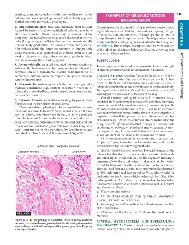
              The classical example of granulomatous inflammation is  M. tuberculosis bovis (bovine strain) used to be common
           the tissue response to tubercle bacilli which is called tubercle  pathogen to human beings during the era of consumption of
           seen in tuberculosis (described below). A fully-developed  unpasteurised milk but presently constitutes a small number
           tubercle is about 1 mm in diameter with central area of  of human cases. Other less common strains included in the
           caseation necrosis, surrounded by epithelioid cells and one  complex are M. africanum (isolated from patients from parts
           to several multinucleated giant cells (commonly Langhans’  of Africa), M. microti, M. pinnipedii and M. canettii. A non-
           type), surrounded at the periphery by lymphocytes and  pathogenic strain, M. smegmatis, is found in the smegma and
           bounded by fibroblasts and fibrous tissue (Fig. 6.19).
                                                               as contaminant in the urine of both men and women.
                                                                  M. tuberculosis hominis is a slender rod-like bacillus,
                                                               0.5 μm by 3  μm, is neutral on Gram staining, and can be
                                                               demonstrated by the following methods:
                                                               1. Acid fast (Ziehl-Neelsen) staining. The acid fastness of the
                                                               tubercle bacilli is due to mycolic acids, cross-linked fatty acids
                                                               and other lipids in the cell wall of the organism making it
                                                               impermeable to the usual stains. It takes up stain by heated
                                                               carbol fuchsin and resists decolourisation by acids and
                                                               alcohols (acid fast and alcohol fast) and can be decolourised
                                                               by 20% sulphuric acid (compared to 5% sulphuric acid for
                                                               declourisation for M. leprae which are less acid fast) (Fig. 6.20).
                                                               False positive AFB staining is seen due to  Nocardia,
                                                               Rhodococcus, Legionella, and some protozoa such as Isospora
                                                               and Cryptosporidium.
                                                               2. Fluorescent dye methods.
                                                               3. Culture of the organism from sputum in Lowenstein-
                                                               Jensen (L.J.) medium for 6 weeks.
                                                               4. Guinea pig inoculation method by subcutaneous injection
                                                               of the organisms.
                                                               5. Molecular methods such as PCR are the most recent
                                                               methods.
           Figure 6.19  Morphology of a tubercle. There is central caseation  ATYPICAL MYCOBACTERIA (NON-TUBERCULOUS
           necrosis, surrounded by elongated epithelioid cells having characteristic
           slipper-shaped nuclei, with interspersed Langhans’ giant cells. Periphery  MYCOBACTERIA). The term atypical mycobacteria or non-
           shows lymphocytes.                                  tuberculous mycobacteria is used for mycobacterial species

