


Page 170 - Textbook of Pathology, 6th Edition
P. 170
154


     SECTION I


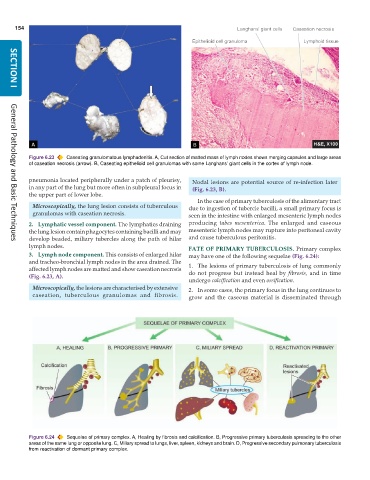
           Figure 6.23  Caseating granulomatous lymphadenitis. A, Cut section of matted mass of lymph nodes shows merging capsules and large areas
           of caseation necrosis (arrow). B, Caseating epithelioid cell granulomas with some Langhans’ giant cells in the cortex of lymph node.

           pneumonia located peripherally under a patch of pleurisy,  Nodal lesions are potential source of re-infection later
           in any part of the lung but more often in subpleural focus in  (Fig. 6.23, B).
           the upper part of lower lobe.
                                                                  In the case of primary tuberculosis of the alimentary tract
            Microscopically, the lung lesion consists of tuberculous  due to ingestion of tubercle bacilli, a small primary focus is
            granulomas with caseation necrosis.                seen in the intestine with enlarged mesenteric lymph nodes

           2. Lymphatic vessel component. The lymphatics draining  producing  tabes mesenterica. The enlarged and caseous
           the lung lesion contain phagocytes containing bacilli and may  mesenteric lymph nodes may rupture into peritoneal cavity
           develop beaded, miliary tubercles along the path of hilar  and cause tuberculous peritonitis.
     General Pathology and Basic Techniques
           lymph nodes.                                        FATE OF PRIMARY TUBERCULOSIS. Primary complex
           3. Lymph node component. This consists of enlarged hilar  may have one of the following sequelae (Fig. 6.24):
           and tracheo-bronchial lymph nodes in the area drained. The
           affected lymph nodes are matted and show caseation necrosis  1. The lesions of primary tuberculosis of lung commonly
           (Fig. 6.23, A).                                     do not progress but instead heal by  fibrosis, and in time
                                                               undergo calcification and even ossification.
            Microscopically, the lesions are characterised by extensive  2. In some cases, the primary focus in the lung continues to
            caseation, tuberculous granulomas and fibrosis.    grow and the caseous material is disseminated through


           Figure 6.24  Sequelae of primary complex. A, Healing by fibrosis and calcification. B, Progressive primary tuberculosis spreading to the other
           areas of the same lung or opposite lung. C, Miliary spread to lungs, liver, spleen, kidneys and brain. D, Progressive secondary pulmonary tuberculosis
           from reactivation of dormant primary complex.

