


Page 176 - Textbook of Pathology, 6th Edition
P. 176
160 1. Upgrading reaction is characterised by increased cell-  The main features in various groups are given below.
           mediated immunity and occurs in patients of borderline  1. Lepromatous leprosy:
           lepromatous (BL) type on treatment who upgrade or shift  The following features characterise lepromatous polar
           towards tuberculoid type.
                                                                 leprosy (Fig. 6.32):
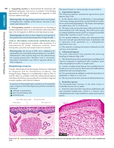
            Histologically, the upgrading reaction shows an increase  i) In the dermis, there is proliferation of macrophages
            of lymphocytes, oedema of the lesions, necrosis in the  with foamy change, particularly around the blood vessels,
            centre and reduced B.I.                              nerves and dermal appendages. The foamy macrophages
                                                                 are called ‘lepra cells’ or Virchow cells.
           2. Downgrading reaction is characterised by lowering of  ii) The lepra cells are heavily laden with acid-fast bacilli
     SECTION I
           cellular immunity and is seen in borderline tuberculoid (BT)  demonstrated with AFB staining. The AFB may be seen
           type who downgrade or shift towards lepromatous type.  as compact globular masses (globi) or arranged in parallel
            Histologically, the lesions show dispersal and spread of  fashion like ‘cigarettes-in-pack’ (see Fig. 6.31).
            the granulomas and increased presence of lepra bacilli.  iii) The dermal infiltrate of lepra cells characteristically
                                                                 does not encroach upon the basal layer of epidermis and
           TYPE II: ERYTHEMA NODOSUM LEPROSUM (ENL).             is separated from epidermis by a subepidermal
           ENL occurs in lepromatous patients after treatment. It is  uninvolved clear zone.
           characterised by tender cutaneous nodules, fever,
           iridocyclitis, synovitis and lymph node involvement.  iv) The epidermis overlying the lesions is thinned out, flat
                                                                 and may even ulcerate.
            Histologically, the lesions in ENL show infiltration by  2. Tuberculoid leprosy:
            neutrophils and eosinophils and prominence of vasculitis.  The polar tuberculoid form presents the following
            Inflammation often extends deep into the subcutaneous  histological features (Fig. 6.33):
            fat causing panniculitis. Bacillary load is increased.
            Secondary amyloidosis may follow repeated attacks of  i) The dermal lesions show granulomas resembling hard
            ENL in leprosy.                                      tubercles composed of epithelioid cells, Langhans’ giant
                                                                 cells and peripheral mantle of lymphocytes.
           Histopathology of Leprosy                             ii) Lesions of tuberculoid leprosy have predilection for
           Usually, skin biopsy from the margin of lesions is submitted  dermal nerves which may be destroyed and infiltrated by
           for diagnosis and for classification of leprosy. The  epithelioid cells and lymphocytes.
           histopathologic diagnosis of multibacillary leprosy like LL  iii) The granulomatous infiltrate erodes the basal layer of
     General Pathology and Basic Techniques
           and BL offers no problem while the indeterminate leprosy  epidermis i.e. there is no clear zone.
           and tuberculoid lesions are paucibacillary and their diagnosis  iv) The lepra bacilli are few and seen in destroyed nerves.
           is made together with clinical evidence.
                                                                 3. Borderline leprosy:
            In general, for histopathologic evaluation in all suspected  The histopathologic features of the three forms of
            cases of leprosy the following broad guidelines should be  borderline leprosy are as under:
            followed:                                            i) Borderline tuberculoid (BT) form shows epithelioid cells
               cell type of granuloma;                           and plentiful lymphocytes. There is a narrow clear
               nerve involvement; and                            subepidermal zone. Lepra bacilli are scanty and found in
               bacterial load.                                   nerves.


           Figure 6.32  Lepromatous leprosy (LL). There is collection of proliferating foam macrophages (lepra cells) in the dermis with a clear subepidermal
           zone.

