


Page 177 - Textbook of Pathology, 6th Edition
P. 177
161


                                                                                                                      CHAPTER 6


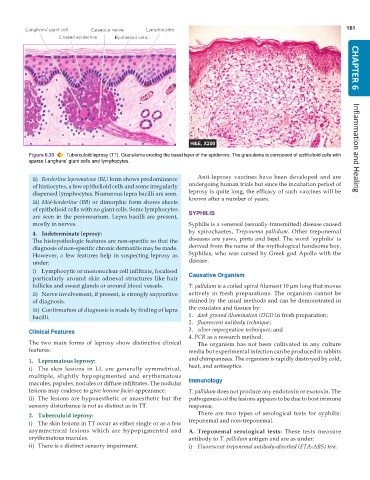
           Figure 6.33  Tuberculoid leprosy (TT). Granuloma eroding the basal layer of the epidermis. The granuloma is composed of epithelioid cells with  Inflammation and Healing
           sparse Langhans’ giant cells and lymphocytes.

            ii) Borderline lepromatous (BL) form shows predominance  Anti-leprosy vaccines have been developed and are
            of histiocytes, a few epithelioid cells and some irregularly  undergoing human trials but since the incubation period of
            dispersed lymphocytes. Numerous lepra bacilli are seen.  leprosy is quite long, the efficacy of such vaccines will be
            iii) Mid-borderline (BB) or dimorphic form shows sheets  known after a number of years.
            of epithelioid cells with no giant cells. Some lymphocytes
            are seen in the peri-neurium. Lepra bacilli are present,  SYPHILIS
            mostly in nerves.                                  Syphilis is a venereal (sexually-transmitted) disease caused

            4. Indeterminate leprosy:                          by spirochaetes,  Treponema pallidum. Other treponemal
            The histopathologic features are non-specific so that the  diseases are yaws, pinta and bejel. The word ‘syphilis’ is
            diagnosis of non-specific chronic dermatitis may be made.  derived from the name of the mythological handsome boy,
            However, a few features help in suspecting leprosy as  Syphilus, who was cursed by Greek god Apollo with the
            under:                                             disease.
            i) Lymphocytic or mononuclear cell infiltrate, focalised
            particularly around skin adnexal structures like hair  Causative Organism
            follicles and sweat glands or around blood vessels.  T. pallidum is a coiled spiral filament 10 μm long that moves
            ii) Nerve involvement, if present, is strongly supportive  actively in fresh preparations. The organism cannot be
            of diagnosis.                                      stained by the usual methods and can be demonstrated in
            iii) Confirmation of diagnosis is made by finding of lepra  the exudates and tissues by:
            bacilli.                                           1. dark ground illumination (DGI) in fresh preparation;
                                                               2. fluorescent antibody technique;
           Clinical Features                                   3. silver impregnation techniques; and
                                                               4. PCR as a research method.
           The two main forms of leprosy show distinctive clinical  The organism has not been cultivated in any culture
           features:                                           media but experimental infection can be produced in rabbits
           1. Lepromatous leprosy:                             and chimpanzees. The organism is rapidly destroyed by cold,
           i) The skin lesions in LL are generally symmetrical,  heat, and antiseptics.
           multiple, slightly hypopigmented and erythematous
           macules, papules, nodules or diffuse infiltrates. The nodular  Immunology
           lesions may coalesce to give leonine facies appearance.  T. pallidum does not produce any endotoxin or exotoxin. The
           ii) The lesions are hypoaesthetic or anaesthetic but the  pathogenesis of the lesions appears to be due to host immune
           sensory disturbance is not as distinct as in TT.    response.
           2. Tuberculoid leprosy:                                There are two types of serological tests for syphilis:
           i) The skin lesions in TT occur as either single or as a few  treponemal and non-treponemal.
           asymmetrical lesions which are hypopigmented and    A. Treponemal serological tests: These tests measure
           erythematous macules.                               antibody to T. pallidum antigen and are as under:
           ii) There is a distinct sensory impairment.         i) Fluorescent treponemal antibody-absorbed (FTA-ABS) test.

