


Page 178 - Textbook of Pathology, 6th Edition
P. 178
162


     SECTION I


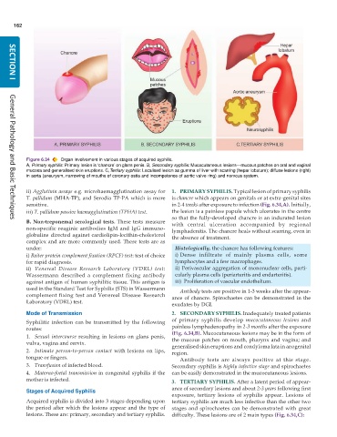
           Figure 6.34  Organ involvement in various stages of acquired syphilis.
           A, Primary syphilis: Primary lesion is ‘chancre’ on glans penis. B, Secondary syphilis: Mucocutaneous lesions—mucous patches on oral and vaginal
           mucosa and generalised skin eruptions. C,Tertiary syphilis: Localised lesion as gumma of liver with scarring (hepar lobatum); diffuse lesions (right)
           in aorta (aneurysm, narrowing of mouths of coronary ostia and incompetence of aortic valve ring) and nervous system.


           ii) Agglutinin assays e.g. microhaemagglutination assay for  1. PRIMARY SYPHILIS. Typical lesion of primary syphilis
           T. pallidum (MHA-TP), and Serodia TP-PA which is more  is chancre which appears on genitals or at extra-genital sites
           sensitive.                                          in 2-4 weeks after exposure to infection (Fig. 6.34,A). Initially,
           iii) T. pallidum passive haemagglutination (TPHA) test.  the lesion is a painless papule which ulcerates in the centre
                                                               so that the fully-developed chancre is an indurated lesion
           B. Non-treponemal serological tests. These tests measure  with central ulceration accompanied by regional
     General Pathology and Basic Techniques
           non-specific reaginic antibodies IgM and IgG immuno-  lymphadenitis. The chancre heals without scarring, even in
           globulins directed against cardiolipin-lecithin-cholesterol  the absence of treatment.
           complex and are more commonly used. These tests are as
           under:                                                Histologically, the chancre has following features:
           i) Reiter protein complement fixation (RPCF) test: test of choice  i) Dense infiltrate of mainly plasma cells, some
           for rapid diagnosis.                                  lymphocytes and a few macrophages.
           ii) Venereal Disease Research Laboratory (VDRL) test:  ii) Perivascular aggregation of mononuclear cells, parti-
           Wassermann described a complement fixing antibody     cularly plasma cells (periarteritis and endarteritis).
           against antigen of human syphilitic tissue. This antigen is  iii)  Proliferation of vascular endothelium.
           used in the Standard Test for Syphilis (STS) in Wassermann  Antibody tests are positive in 1-3 weeks after the appear-
           complement fixing test and Venereal Disease Research  ance of chancre. Spirochaetes can be demonstrated in the
           Laboratory (VDRL) test.
                                                               exudates by DGI.
           Mode of Transmission                                2. SECONDARY SYPHILIS. Inadequately treated patients
           Syphilitic infection can be transmitted by the following  of primary syphilis develop  mucocutaneous lesions and
           routes:                                             painless lymphadenopathy in 2-3 months after the exposure
           1. Sexual intercourse resulting in lesions on glans penis,  (Fig. 6.34,B). Mucocutaneous lesions may be in the form of
                                                               the mucous patches on mouth, pharynx and vagina; and
           vulva, vagina and cervix.                           generalised skin eruptions and condyloma lata in anogenital
           2. Intimate person-to-person contact with lesions on lips,  region.
           tongue or fingers.                                     Antibody tests are always positive at this stage.
           3. Transfusion of infected blood.                   Secondary syphilis is highly infective stage and spirochaetes
           4. Materno-foetal transmission in congenital syphilis if the  can be easily demonstrated in the mucocutaneous lesions.
           mother is infected.                                 3. TERTIARY SYPHILIS. After a latent period of appear-
                                                               ance of secondary lesions and about 2-3 years following first
           Stages of Acquired Syphilis
                                                               exposure, tertiary lesions of syphilis appear. Lesions of
           Acquired syphilis is divided into 3 stages depending upon  tertiary syphilis are much less infective than the other two
           the period after which the lesions appear and the type of  stages and spirochaetes can be demonstrated with great
           lesions. These are: primary, secondary and tertiary syphilis.  difficulty. These lesions are of 2 main types (Fig. 6.34,C):

