


Page 179 - Textbook of Pathology, 6th Edition
P. 179
ii) The characteristic ‘Hutchinson’s teeth’ which are small,  163
                                                               widely spaced, peg-shaped permanent teeth.
                                                               iii) Mucocutaneous lesions of acquired secondary syphilis.
                                                               iv) Bony lesions like epiphysitis and periostitis.
                                                               v) Interstitial keratitis with corneal opacity.
                                                               vi) Diffuse fibrosis in the liver.
                                                               vii)Interstitial fibrosis of lungs.                    CHAPTER 6
                                                               viii) If the foetus with congenital syphilis is born dead, it is
                                                               premature, with macerated skin, enlarged spleen and liver,
                                                               and with syphilitic epiphysitis.
                                                                 Histologically, the basic morphology of lesions in syphilis
                                                                 is seen in all the affected organs: perivascular plasma cell
                                                                 rich inflammatory infiltrate and endothelial cell
                                                                 proliferation. Many spirochaetes can be demonstrated in
                                                                 involved tissues.

                                                               ACTINOMYCOSIS                                          Inflammation and Healing
                                                               Actinomycosis is a chronic suppurative disease caused by
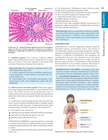
           Figure 6.35  Typical microscopic appearance in the case of syphilitic
           gumma of the liver. Central coagulative necrosis is surrounded by  anaerobic bacteria,  Actinomycetes israelii. The disease is
           palisades of macrophages and plasma cells marginated peripherally by  conventionally included in mycology though the causative
           fibroblasts.                                        organism is filamentous bacteria and not true fungus. The
                                                               disease is worldwide in distribution. The organisms are
           i) Syphilitic gumma. It is a solitary, localised, rubbery  commensals in the oral cavity, alimentary tract and vagina.
           lesion with central necrosis, seen in organs like liver, testis,  The infection is always endogeneous in origin and not by
           bone and brain. In liver, the gumma is associated with  person-to-person contact. The organisms invade, proliferate
           scarring of hepatic parenchyma (hepar lobatum).     and disseminate in favourable conditions like break in
                                                               mucocutaneous continuity, some underlying disease etc.
            Histologically, the structure of gumma shows the
            following features (Fig. 6.35):                      MORPHOLOGIC FEATURES. Depending upon the
            a) Central coagulative necrosis resembling caseation but  anatomic location of lesions, actinomycosis is of 4 types:
            is less destructive so that outlines of necrosed cells can  cervicofacial, thoracic, abdominal, and pelvic (Fig. 6.36).
            still be faintly seen.                               1. Cervicofacial actinomycosis. This is the commonest
            b) Surrounding zone of palisaded macrophages with    form (60%) and has the best prognosis. The infection enters
            many plasma cells, some lymphocytes, giant cells and  from tonsils, carious teeth, periodontal disease or trauma
            fibroblasts.                                         following tooth extraction. Initially, a firm swelling
                                                                 develops in the lower jaw (‘lumpy jaw’). In time, the mass
           ii) Diffuse lesions of tertiary syphilis. The lesions appear  breaks down and abscesses and sinuses are formed. The
           following widespread dissemination of spirochaetes in the  discharging pus contains typical tiny yellow sulphur
           body. The diffuse lesions are predominantly seen in
           cardiovascular and nervous systems which are described in
           detail later in the relevant chapters. Briefly, these lesions are
           as under:
           a) Cardiovascular syphilis mainly involves thoracic aorta. The
           wall of aorta is weakened and dilated due to syphilitic aortitis
           and results in aortic aneurysm, incompetence of aortic valve
           and narrowing of mouths of coronary ostia (Chapter 15).
           b) Neurosyphilis may manifest as:
              meningovascular syphilis affecting chiefly the meninges;
              tabes dorsalis affecting the spinal cord; and
              general paresis affecting the brain.

           CONGENITAL SYPHILIS. Congenital syphilis may develop
           in a foetus of more than 16 weeks gestation who is exposed
           to maternal spirochaetaemia. The major morphologic features
           as under:
           i) Saddle-shaped nose deformity due to destruction of bridge
           of the nose.                                        Figure 6.36  Actinomycosis, sites and routes of infection.

