


Page 315 - Textbook of Pathology, 6th Edition
P. 315
299


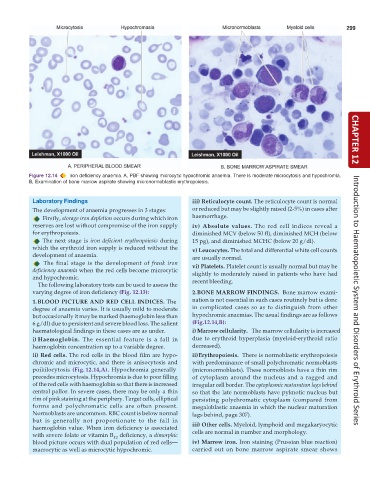
           Figure 12.14  Iron deficiency anaemia. A, PBF showing microcytic hypochromic anaemia. There is moderate microcytosis and hypochromia.  CHAPTER 12
           B, Examination of bone marrow aspirate showing micronormoblastic erythropoiesis.


            Laboratory Findings                                  iii) Reticulocyte count. The reticulocyte count is normal
            The development of anaemia progresses in 3 stages:   or reduced but may be slightly raised (2-5%) in cases after
               Firstly, storage iron depletion occurs during which iron  haemorrhage.
            reserves are lost without compromise of the iron supply  iv)  Absolute  values. The red cell indices reveal a
            for erythropoiesis.                                  diminished MCV (below 50 fl), diminished MCH (below
               The next stage is  iron deficient  erythropoiesis during  15 pg), and diminished MCHC (below 20 g/dl).
            which the erythroid iron supply is reduced without the  v) Leucocytes. The total and differential white cell counts
            development of anaemia.                              are usually normal.
               The final stage is the development of  frank iron  vi) Platelets. Platelet count is usually normal but may be
            deficiency anaemia when the red cells become microcytic  slightly to moderately raised in patients who have had
            and hypochromic.                                     recent bleeding.
              The following laboratory tests can be used to assess the
            varying degree of iron deficiency (Fig. 12.13):      2.BONE MARROW FINDINGS.  Bone marrow exami-
            1.BLOOD PICTURE AND RED CELL INDICES. The            nation is not essential in such cases routinely but is done  Introduction to Haematopoietic System and Disorders of Erythroid Series
            degree of anaemia varies. It is usually mild to moderate  in complicated cases so as to distinguish from other
            but occasionally it may be marked (haemoglobin less than  hypochromic anaemias. The usual findings are as follows
            6 g/dl) due to persistent and severe blood loss. The salient  (Fig.12.14,B):
            haematological findings in these cases are as under.  i) Marrow cellularity.  The marrow cellularity is increased
            i) Haemoglobin. The essential feature is a fall in   due to erythroid hyperplasia (myeloid-erythroid ratio
            haemoglobin concentration up to a variable degree.   decreased).
            ii) Red cells. The red cells in the blood film are hypo-  ii)Erythropoiesis.  There is normoblastic erythropoiesis
            chromic and microcytic, and there is anisocytosis and  with predominance of small polychromatic normoblasts
            poikilocytosis  (Fig. 12.14,A). Hypochromia generally  (micronormoblasts). These normoblasts have a thin rim
            precedes microcytosis. Hypochromia is due to poor filling  of cytoplasm around the nucleus and a ragged and
            of the red cells with haemoglobin so that there is increased  irregular cell border. The cytoplasmic maturation lags behind
            central pallor. In severe cases, there may be only a thin  so that the late normoblasts have pyknotic nucleus but
            rim of pink staining at the periphery. Target cells, elliptical  persisting polychromatic cytoplasm (compared from
            forms and polychromatic cells are often present.     megaloblastic anaemia in which the nuclear maturation
            Normoblasts are uncommon. RBC count is below normal  lags behind, page 307).
            but is generally not proportionate to the fall in
            haemoglobin value. When iron deficiency is associated  iii) Other cells. Myeloid, lymphoid and megakaryocytic
                                                                 cells are normal in number and morphology.
            with severe folate or vitamin B  deficiency, a dimorphic
                                       12
            blood picture occurs with dual population of red cells—  iv) Marrow iron. Iron staining (Prussian blue reaction)
            macrocytic as well as microcytic hypochromic.        carried out on bone marrow aspirate smear shows

