


Page 317 - Textbook of Pathology, 6th Edition
P. 317
spontaneously in middle-aged and older individuals of both  301
                                                               sexes. The disorder has its pathogenesis in disturbed growth
                                                               and maturation of erythroid precursors at the level of
                                                               haematopoietic stem cell, possibly due to reduced activity
                                                               of the enzyme, ALA synthetase. The anaemia is of moderate
                                                               to severe degree and appears insidiously. The bone marrow
                                                               cells commonly show chromosomal abnormalities, neutro-
                                                               penia and thrombocytopenia with associated bleeding
                                                               diathesis. The spleen and liver may be either normal or mildly
                                                               enlarged, while the lymph nodes are not enlarged. Unlike
                                                               other types of sideroblastic anaemia, this type is regarded as
                                                               a myelodysplastic disorder in the FAB (French-American-
                                                               British) classification and thus, can be a preleukaemic
                                                               disorder (page 361). About 10% of individuals with refractory
                                                               acquired sideroblastic anaemia develop acute myelogenous
                                                               leukaemia.                                             CHAPTER 12
                                                               B. Secondary acquired  sideroblastic  anaemia.  Acquired
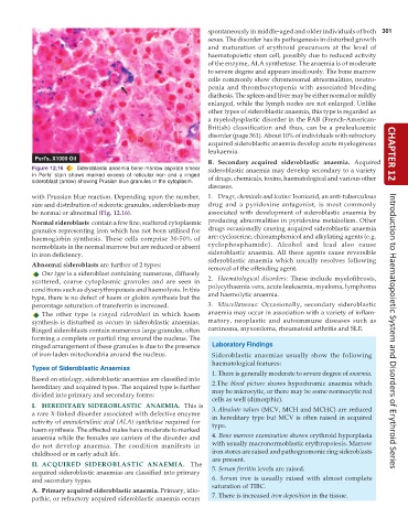
           Figure 12.16  Sideroblastic anaemia bone marrow aspirate smear  sideroblastic anaemia may develop secondary to a variety
           in Perls’ stain shows marked excess of reticular iron and a ringed  of drugs, chemicals, toxins, haematological and various other
           sideroblast (arrow) showing Prusian blue granules in the cytoplasm.
                                                               diseases.
           with Prussian blue reaction. Depending upon the number,  1. Drugs, chemicals and toxins: Isoniazid, an anti-tuberculous
           size and distribution of siderotic granules, sideroblasts may  drug and a pyridoxine antagonist, is most commonly
           be normal or abnormal (Fig. 12.16).                 associated with development of sideroblastic anaemia by
           Normal sideroblasts  contain a few fine, scattered cytoplasmic  producing abnormalities in pyridoxine metabolism. Other
           granules representing iron which has not been utilised for  drugs occasionally causing acquired sideroblastic anaemia
           haemoglobin synthesis. These cells comprise 30-50% of  are: cycloserine, chloramphenicol and alkylating agents (e.g.
           normoblasts in the normal marrow but are reduced or absent  cyclophosphamide). Alcohol and lead also cause
           in iron deficiency.                                 sideroblastic anaemia. All these agents cause reversible
                                                               sideroblastic anaemia which usually resolves following
           Abnormal sideroblasts are further of 2 types:       removal of the offending agent.
              One type is a sideroblast containing numerous, diffusely
           scattered, coarse cytoplasmic granules and are seen in  2. Haematological disorders: These include myelofibrosis,
           conditions such as dyserythropoiesis and haemolysis. In this  polycythaemia vera, acute leukaemia, myeloma, lymphoma
           type, there is no defect of haem or globin synthesis but the  and haemolytic anaemia.
           percentage saturation of transferrin is increased.  3. Miscellaneous: Occasionally, secondary sideroblastic
              The other type is  ringed sideroblast in which haem  anaemia may occur in association with a variety of inflam-
           synthesis is disturbed as occurs in sideroblastic anaemias.  matory, neoplastic and autoimmune diseases such as
           Ringed sideroblasts contain numerous large granules, often  carcinoma, myxoedema, rheumatoid arthritis and SLE.  Introduction to Haematopoietic System and Disorders of Erythroid Series
           forming a complete or partial ring around the nucleus. The
           ringed arrangement of these granules is due to the presence  Laboratory Findings
           of iron-laden mitochondria around the nucleus.        Sideroblastic anaemias usually show the following
                                                                 haematological features:
           Types of Sideroblastic Anaemias
                                                                 1. There is generally moderate to severe degree of anaemia.
           Based on etiology, sideroblastic anaemias are classified into
           hereditary and acquired types. The acquired type is further  2.The  blood picture shows hypochromic anaemia which
           divided into primary and secondary forms:             may be microcytic, or there may be some normocytic red
                                                                 cells as well (dimorphic).
           I. HEREDITARY SIDEROBLASTIC  ANAEMIA.  This is        3. Absolute values (MCV, MCH and MCHC) are reduced
           a rare X-linked disorder associated with defective enzyme  in hereditary type but MCV is often raised in acquired
           activity of aminolevulinic acid (ALA) synthetase required for  type.
           haem synthesis. The affected males have moderate to marked
           anaemia while the females are carriers of the disorder and  4. Bone marrow examination shows erythroid hyperplasia
           do not develop anaemia. The condition manifests in    with usually macronormoblastic erythropoiesis. Marrow
           childhood or in early adult life.                     iron stores are raised and pathognomonic ring sideroblasts
                                                                 are present.
           II. ACQUIRED  SIDEROBLASTIC ANAEMIA.   The
           acquired sideroblastic anaemias are classified into primary  5. Serum ferritin levels are raised.
           and secondary types.                                  6.  Serum iron is usually raised with almost complete
                                                                 saturation of TIBC.
           A. Primary acquired sideroblastic anaemia. Primary, idio-
           pathic, or refractory acquired sideroblastic anaemia occurs  7. There is increased iron deposition in the tissue.

