


Page 337 - Textbook of Pathology, 6th Edition
P. 337
321
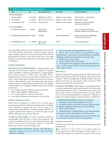
TABLE 12.12: Classification of Thalassaemias.
Type Hb Hb-electrophoresis Genotype Clinical Syndrome
α-THALASSAEMIAS
1. Hydrops foetalis 3-10 gm/dl Hb Barts (g 4 ) (100%) Deletion of four α-genes Fatal in utero or in early infancy
2. Hb-H disease 2-12 gm/dl HbF (10%), HbH (2-4%) Deletion of three α-genes Haemolytic anaemia
3. α-Thalassaemia trait 10-14 gm/dl Almost normal Deletion of two α-genes Microcytic hypochromic blood
picture but no anaemia
β-THALASSAEMIAS
1. β-Thalassaemia major < 5 gm/dl HbA (0-50%), β thal /β thal Severe congenital haemolytic
HbF(50-98%) anaemia, requires blood transfusions
2. β-Thalassaemia intermedia 5-10 gm/dl Variable Multiple mechanisms Severe anaemia, but regular blood
transfusions not required
A
3. β-Thalassaemia minor 10-12 gm/dl HbA 2 (4-9%) β /β thal Usually asymptomatic CHAPTER 12
HbF (1-5%)
are irreparably damaged and are phagocytosed by the RE 1. Severe anaemia (haemoglobin below 6g/dl).
cells of the spleen and the liver causing anaemia, hepato- 2. Blood film show marked anisopoikilocytosis,
splenomegaly, and excess of tissue iron stores. Patients with hypochromia, microcytosis, polychromasia, basophilic
β-thalassaemia minor, on the other hand, have very mild stippling, numerous normoblasts and target cells.
ineffective erythropoiesis, haemolysis and shortening of red 3. Reticulocyte count is high.
cell lifespan. 4. Serum bilirubin level is elevated.
5. Haemoglobin electrophoresis shows 80-90% Hb-Bart’s
α α α α α-THALASSAEMIA and a small amount of Hb-H and Hb-Portland but no
MOLECULAR PATHOGENESIS. α-thalassaemias are HbA, HbA or HbF.
2
disorders in which there is defective synthesis of α-globin HbH Disease
chains resulting in depressed production of haemoglobins
that contain α-chains i.e. HbA, HbA and HbF. The α- Deletion of three α-chain genes produces HbH which is a β-
2
thalassaemias are most commonly due to deletion of one or globin chain tetramer (β ) and markedly impaired α-chain
4
more of the α-chain genes located on short arm of chromo- synthesis. HbH is precipitated as Heinz bodies within the
some 16. Since there is a pair of α-chain genes, the clinical affected red cells. An elongated α-chain variant of HbH
manifestations of α-thalassaemia depend upon the number disease is termed Hb Constant Spring.
of genes deleted. Accordingly, α-thalassaemias are classified CLINICAL FEATURES. HbH disease is generally present
into 4 types: as a well-compensated haemolytic anaemia. The features are
1. Four α-gene deletion: Hb Bart’s hydrops foetalis. intermediate between that of β-thalassaemia minor and Introduction to Haematopoietic System and Disorders of Erythroid Series
2. Three α-gene deletion: HbH disease. major. The severity of anaemia fluctuates and may fall to
3. Two α-gene deletion: α-thalassaemia trait. very low levels during pregnancy or infections. Majority of
4. One α-gene deletion: α-thalassaemia trait (carrier). patients have splenomegaly and may develop cholelithiasis.
Hb Bart’s Hydrops Foetalis LABORATORY FINDINGS. These are as follows:
When there is deletion of all the four α-chain genes 1. Moderate anaemia (haemoglobin 8-9 g/dl).
(homozygous state) it results in total suppression of α-globin 2. Blood film shows severe microcytosis, hypochromia,
chain synthesis causing the most severe form of α- basophilic stippling, target cells and normoblasts.
thalassaemia called Hb Bart’s hydrops foetalis. Hb Bart’s is 3. Mild reticulocytosis.
a gamma globin chain tetramer (γ ) which has high oxygen 4. HbH inclusions as Heinz bodies can be demonstrated
4
affinity leading to severe tissue hypoxia. in mature red cells with brilliant cresyl blue stain.
5. Haemoglobin electrophoresis shows 2-4% HbH and
CLINICAL FEATURES. Hb Bart’s hydrops foetalis is the remainder consists of HbA, HbA and HbF.
incompatible with life due to severe tissue hypoxia. The 2
condition is either fatal in utero or the infant dies shortly after α α α α α-Thalassaemia Trait
birth. If born alive, the features similar to severe Rh α-thalassaemia trait may occur by the following molecular
haemolytic disease are present (page 340).
pathogenesis:
LABORATORY FINDINGS. Infants with Hb Bart’s By deletion of two of the four α-chain genes in
hydrops foetalis born alive may have the following homozygous form called homozygous α-thalassaemia, or in
laboratory findings: double heterozygous form termed heterozygous α-thalassaemia.

