


Page 338 - Textbook of Pathology, 6th Edition
P. 338
322     By deletion of a single α-chain gene causing heterozygous
           α-thalassaemia trait called heterozygous α-thalassaemia.
           CLINICAL FEATURES.  α-thalassaemia trait due to two
           α-chain gene deletion is asymptomatic. It is suspected in a
           patient of refractory microcytic hypochromic anaemia in
           whom iron deficiency and β-thalassaemia minor have been
           excluded and the patient belongs to the high-risk ethnic
           group. One gene deletion  α-thalassaemia trait is a silent
           carrier state.
            LABORATORY FINDINGS.  The patients of α-thalas-
            saemia trait may have the following haematological
            findings:
            1. Haemoglobin level normal or mildly reduced.
            2. Blood film shows microcytic and hypochromic red cell
            morphology but no evidence of haemolysis or anaemia.
            3. MCV, MCH and MCHC may be slightly reduced.
            4. Haemoglobin electrophoresis reveals small amount of
     SECTION II
            Hb-Bart’s in neonatal period (1-2% in α-thalassaemia 2
            and 5-6% in α-thalassaemia 1) which gradually disappears
            by adult life. HbA  is either normal or slightly decreased
                           2
            (contrary to the elevated HBA  levels in β-thalassaemia
                                      2
            trait).
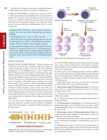
                                                               Figure 12.31  Pathogenesis of β-thalassaemia major.
           β β β β β-THALASSAEMIAS
                                                               ii) Translation defect: Mutation in the coding sequence causing
           MOLECULAR  PATHOGENESIS.   β-thalassaemias are      stop codon (chain termination) interrupting  β-globin
           caused by decreased rate of β-chain synthesis resulting in  messenger RNA. This would result in no synthesis of β-globin
           reduced formation of HbA in the red cells. The molecular  chain i.e. β° thalassaemia.
           pathogenesis of the β-thalassaemias is more complex than  iii) mRNA splicing defect: Mutation leads to defective mRNA
           that of α-thalassaemias. In contrast to α-thalassaemia, gene  processing forming abnormal mRNA that is degraded in the
           deletion rarely ever causes β-thalassaemia and is only seen  nucleus. Depending upon whether part of splice site remains
           in an entity called hereditary persistence of foetal haemoglobin  intact or is totally degraded, it may result in β  thalassaemia
                                                                                                       +
           (HPFH). Instead, most of β-thalassaemias arise from different  or β° thalassaemia.
           types of mutations of β−globin gene resulting from single  Depending upon the extent of reduction in  β-chain
           base changes. The symbol β° is used to indicate the complete  synthesis, there are 3 types of β-thalassaemia:
                                               +
           absence of β-globin chain synthesis while β  denotes partial
           synthesis of the  β-globin chains. More than 100 such  1. Homozygous form: β-Thalassaemia major. It is the most
           mutations have been described affecting the preferred sites  severe form of congenital haemolytic anaemia. It is further
           in the coding sequences e.g. in promoter region, termination  of 2 types (Fig. 12.31):
           region, splice junctions, exons, introns. Some of the important  i) β° thalassaemia major characterised by complete absence
     Haematology and Lymphoreticular Tissues
           ones having effects on β-globin chain synthesis are as under  of β-chain synthesis.
                                                                   +
           (Fig. 12.30):                                       ii) β  thalassaemia major having incomplete suppression of
           i)  Transcription defect: Mutation affecting transcriptional  β-chain synthesis.
           promoter sequence causing reduced synthesis of β-globin  2. β-Thalassaemia  intermedia: It is  β-thalassaemia of
           chain. Hence the result is partially preserved synthesis i.e.  intermediate degree of severity that does not require regular
           β  thalassaemia.                                    blood transfusions. These cases are genetically heterozygous
            +
                                                                        +
                                                               (β°/β or β /β).
                                                               3. Heterozygous form: β-Thalassaemia minor (trait). It is
                                                               a mild asymptomatic condition in which there is moderate
                                                               suppression of β-chain synthesis.
                                                                  Besides β-thalassaemia minor, a few uncommon globin
                                                               chain combinations resulting in β-thalassaemia trait are as
                                                               under:
                                                               i) δβ-thalassaemia minor in which there is total absence of
                                                               both β and δ chain synthesis and is characterised by elevated
           Figure 12.30  Schematic representation of sites of β-globin gene  HbF level but unlike β-thalassaemia minor there is normal
           mutation in chromosome 11 giving rise to β-thalassaemia.  or reduced HbA  level.
                                                                             2

