


Page 443 - Textbook of Pathology, 6th Edition
P. 443
ISCHAEMIC HEART DISEASE                                                                         427
           Ischaemic heart disease (IHD) is defined as acute or chronic
           form of cardiac disability arising from imbalance between
           the myocardial supply and demand for oxygenated blood.
           Since narrowing or obstruction of the coronary arterial
           system is the most common cause of myocardial anoxia, the
           alternate term  ‘coronary artery disease (CAD)’ is used
           synonymously with IHD. IHD or CAD is the leading cause
           of death in most developed countries (about one-third of all
           deaths) and somewhat low incidence is observed in the
           developing countries. Men develop IHD earlier than women
           and death rates are also slightly higher for men than for
           women until the menopause. As per rising trends of IHD
           worldwide, it is estimated that by the year 2020 it would
           become the most common cause of death throughout world.

           ETIOPATHOGENESIS
           IHD is invariably caused by disease affecting the coronary
           arteries, the most prevalent being atherosclerosis accounting
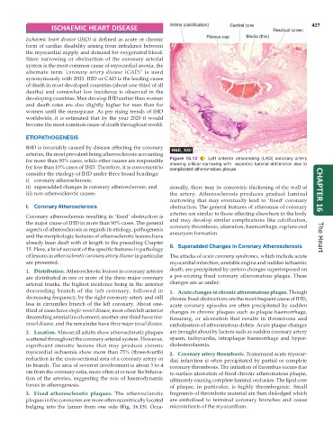
           for more than 90% cases, while other causes are responsible  Figure 16.13  Left anterior descending (LAD) coronary artery
           for less than 10% cases of IHD. Therefore, it is convenient to  showing critical narrowing with  eccentric luminal obliteration due to
                                                               complicated atheromatous plaque.
           consider the etiology of IHD under three broad headings:
           i) coronary atherosclerosis;
           ii) superadded changes in coronary atherosclerosis; and  sionally, there may be concentric thickening of the wall of
           iii) non-atherosclerotic causes.                    the artery. Atherosclerosis produces gradual luminal   CHAPTER 16
                                                               narrowing that may eventually lead to ‘fixed’ coronary
           I.  Coronary Atherosclerosis                        obstruction. The general features of atheromas of coronary
                                                               arteries are similar to those affecting elsewhere in the body
           Coronary atherosclerosis resulting in ‘fixed’ obstruction is
           the major cause of IHD in more than 90% cases. The general  and may develop similar complications like calcification,
           aspects of atherosclerosis as regards its etiology, pathogenesis  coronary thrombosis, ulceration, haemorrhage, rupture and
           and the morphologic features of atherosclerotic lesions have  aneurysm formation.                          The Heart
           already been dealt with at length in the preceding Chapter
           15. Here, a brief account of the specific features in pathology  II. Superadded Changes in Coronary Atherosclerosis
           of lesions in atherosclerotic coronary artery disease in particular  The attacks of acute coronary syndromes, which include acute
           are presented.                                      myocardial infarction, unstable angina and sudden ischaemic
           1. Distribution. Atherosclerotic lesions in coronary arteries  death, are precipitated by certain changes superimposed on
           are distributed in one or more of the three major coronary  a pre-existing fixed coronary atheromatous plaque. These
           arterial trunks, the highest incidence being in the anterior  changes are as under:
           descending branch of the left coronary, followed in  1. Acute changes in chronic atheromatous plaque. Though
           decreasing frequency, by the right coronary artery and still  chronic fixed obstructions are the most frequent cause of IHD,
           less in circumflex branch of the left coronary. About one-  acute coronary episodes are often precipitated by sudden
           third of cases have single-vessel disease, most often left anterior  changes in chronic plaques such as plaque haemorrhage,
           descending arterial involvement; another one-third have two-  fissuring, or ulceration that results in thrombosis and
           vessel disease, and the remainder have three major vessel disease.  embolisation of atheromatous debris. Acute plaque changes
           2. Location. Almost all adults show atherosclerotic plaques  are brought about by factors such as sudden coronary artery
           scattered throughout the coronary arterial system. However,  spasm, tachycardia, intraplaque haemorrhage and hyper-
           significant stenotic lesions that may produce chronic  cholesterolaemia.
           myocardial ischaemia show more than 75% (three-fourth)  2. Coronary artery thrombosis. Transmural acute myocar-
           reduction in the cross-sectional area of a coronary artery or  dial infarction is often precipitated by partial or complete
           its branch. The area of severest involvement is about 3 to 4  coronary thrombosis. The initiation of thrombus occurs due
           cm from the coronary ostia, more often at or near the bifurca-  to surface ulceration of fixed chronic atheromatous plaque,
           tion of the arteries, suggesting the role of haemodynamic  ultimately causing complete luminal occlusion. The lipid core
           forces in atherogenesis.                            of plaque, in particular, is highly thrombogenic. Small
           3. Fixed atherosclerotic plaques.  The atherosclerotic  fragments of thrombotic material are then dislodged which
           plaques in the coronaries are more often eccentrically located  are embolised to terminal coronary branches and cause
           bulging into the lumen from one side (Fig. 16.13). Occa-  microinfarcts of the myocardium.

