


Page 444 - Textbook of Pathology, 6th Edition
P. 444
428
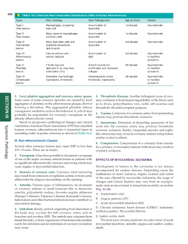
TABLE 16.2: American Heart Association Classification (1995) of Human Atherosclerosis.
Types Main Histology Main Pathogenesis Age at Onset Clinical
Type I: Macrophages, occasional Accumulation of 1st decade Asymptomatic
Initial lesions foam cell lipoprotein
Type II: Many layers of macrophages Accumulation of 1st decade Asymptomatic
Fatty streaks and foam cells lipoprotein
Type III: Many lipid-laden cells and Accumulation of 3rd decade Asymptomatic
Intermediate scattered extracellular lipoprotein
lesions lipid droplets
Type IV: Intra-as well as extra- Accumulation of 3rd decade Asymptomatic
Atheromatous cellular lipid pool lipid or manifest
lesions symptoms
Type V: Fibrotic cap and Smooth muscle cell 4th decade Asymptomatic
Fibrofatty lipid core (V a), may have proliferation and increased or manifest
lesions calcification (V b) collagen symptoms
Type VI: Ulceration, haemorrhage, Haemodynamic stress, 4th decade Asymptomatic
Complicated haematoma, thrombosis thrombosis, haematoma or manifest
lesions symptoms
3. Local platelet aggregation and coronary artery spasm. 5. Thrombotic diseases. Another infrequent cause of coro-
Some cases of acute coronary episodes are caused by local nary occlusion is from hypercoagulability of the blood such
aggregates of platelets on the atheromatous plaque, short of as in shock, polycythaemia vera, sickle cell anaemia and
forming a thrombus. The aggregated platelets release thrombotic thrombocytopenic purpura.
vasospasmic mediators such as thromboxane A which may
2
probably be responsible for coronary vasospasm in the 6. Trauma. Contusion of a coronary artery from penetrating
already atherosclerotic vessel. injuries may produce thrombotic occlusion.
SECTION III
Based on progressive pathological changes and clinical 7. Aneurysms. Extension of dissecting aneurysm of the
correlation, American Heart Association (1995) has classified aorta into the coronary artery may produce thrombotic
human coronary atherosclerosis into 6 sequential types in coronary occlusion. Rarely, congenital, mycotic and syphi-
ascending order of grades of lesions as shown in Table 16.2. litic aneurysms may occur in coronary arteries and produce
similar occlusive effects.
III. Non-atherosclerotic Causes
8. Compression. Compression of a coronary from outside
Several other coronary lesions may cause IHD in less than by a primary or secondary tumour of the heart may result in
10% of cases. These are as under: coronary occlusion.
1. Vasospasm. It has been possible to document vasospasm
of one of the major coronary arterial trunks in patients with EFFECTS OF MYOCARDIAL ISCHAEMIA
Systemic Pathology
no significant atherosclerotic coronary narrowing which may
cause angina or myocardial infarction. Development of lesions in the coronaries is not always
accompanied by cardiac disease. Depending upon the
2. Stenosis of coronary ostia. Coronary ostial narrowing suddenness of onset, duration, degree, location and extent
may result from extension of syphilitic aortitis or from aortic of the area affected by myocardial ischaemia, the range of
atherosclerotic plaques encroaching on the opening.
changes and clinical features may vary from an asympto-
3. Arteritis. Various types of inflammatory involvements matic state at one extreme to immediate mortality at another
of coronary arteries or small branches like in rheumatic (Fig. 16.14):
arteritis, polyarteritis nodosa, thrombo-angiitis obliterans A. Asymptomatic state
(Buerger’s disease), Takayasu’s disease, Kawasaki’s disease,
tuberculosis and other bacterial infections may contribute to B. Angina pectoris (AP)
myocardial damage. C. Acute myocardial infarction (MI)
D. Chronic ischaemic heart disease (CIHD)/ Ischaemic
4. Embolism. Rarely, emboli originating from elsewhere in
the body may occlude the left coronary artery and its cardiomyopathy/ Myocardial fibrosis
branches and produce IHD. The emboli may originate from E. Sudden cardiac death
bland thrombi, or from vegetations of bacterial endocarditis; The term acute coronary syndromes include a triad of acute
rarely fat embolism and air embolism of coronary circulation myocardial infarction, unstable angina and sudden cardiac
may occur. death.

