


Page 450 - Textbook of Pathology, 6th Edition
P. 450
434


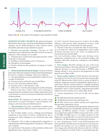
           Figure 16.20  Some common ECG changes in acute myocardial infarction.


           CHANGES IN EARLY INFARCTS. By special techniques    iv) Shock: Systolic blood pressure is below 80 mmHg;
           like electron microscopy, chemical and histochemical studies,  lethargy, cold clammy limbs, peripheral cyanosis, weak
           changes can be demonstrated in early infarcts before  pulse, tachycardia or bradycardia are often present.
           detectable light microscopic alterations appear.    v) Oliguria: Urine flow is usually less than 20 ml per hour.
           1. Electron microscopic changes.  Changes by EM     vi) Low grade fever: Mild rise in temperature occurs within
           examination are evident in less than half an hour on onset of  24 hours and lasts up to one week, accompanied by
           infarction. These changes are as under:             leucocytosis and elevated ESR.
           i) Disappearance of perinuclear glycogen granules within  vii)  Acute pulmonary oedema: Some cases develop severe
           5 minutes of ischaemia.                             pulmonary congestion due to left ventricular failure and
           ii) Swelling of mitochondria in 20 to 30 minutes.   develop suffocation, dyspnoea, orthopnoea and bubbling
           iii) Disruption of sarcolemma.                      respiration.
           iv) Nuclear alterations like peripheral clumping of nuclear  2. ECG changes.  The ECG changes are one of the most
           chromatin.                                          important parameters. Most characteristic ECG change is ST
     SECTION III
                                                               segment elevation in acute MI (termed as STEMI); other
           2. Chemical and histochemical changes. Analysis of tissues  changes inlcude T wave inversion and appearance of wide
           from early infarcts by chemical and histochemical techniques  deep Q waves (Fig. 16.20).
           has shown a number of findings. These are as follows:  3. Serum cardiac markers. Certain proteins and enzymes
           i) Glycogen depletion in myocardial fibres within 30 to 60  are released into the blood from necrotic heart muscle after
           minutes of infarction.                              acute MI. Measurement of their levels in serum is helpful in
           ii) Increase in lactic acid in the myocardial fibres.  making a diagnosis and plan management. Rapid assay of
                     +
           iii) Loss of K  from the ischaemic fibres.          some more specific cardiac proteins is available rendering
                          +
           iv) Increase of Na  in the ischaemic cells.         the estimation of non-specific estimation of SGOT of historical
                       ++
           v) Influx of Ca  into the cells causing irreversible cell injury.  importance only in current practice. Important myocardial
              Based on the above observations and on leakage of  markers in use nowadays are as under (Fig. 16.21):
     Systemic Pathology
           enzymes from the ischaemic myocardium, alterations in the
           concentrations of various enzymes are detected in the blood  i) Creatine phosphokinase  (CK) and CK-MB: CK has three
           of these patients.                                  forms—
                                                                  CK-MM derived from skeletal muscle;
           DIAGNOSIS. The diagnosis of acute MI is made on the    CK-BB derived from brain and lungs; and
           observations of 3 types of features—clinical features, ECG  CK-MB, mainly from cardiac muscles and insignificant
           changes, and serum enzyme determinations.           amount from extracardiac tissue.
           1. Clinical features. Typically, acute MI has a sudden onset.  Thus total CK estimation lacks specificity while elevation
           The following clinical features usually characterise a case of  of CK-MB isoenzyme is considerably specific for myocardial
           acute MI.                                           damage. CK-MB has further 2 forms—CK-MB2 is the
           i) Pain: Usually sudden, severe, crushing and prolonged,  myocardial form while CK-MB1 is extracardiac form. A ratio
           substernal or precordial in location, unrelieved by rest or  of CK-MB2: CK-MB1 above 1.5 is highly sensitive for the
           nitroglycerin, often radiating to one or both the arms, neck  diagnosis of acute MI after 4-6 hours of onset of myocardial
           and back.                                           ischaemia. CK-MB disappears from blood by 48 hours.
           ii) Indigestion: Pain is often accompanied by epigastric or  ii) Lactic dehydrogenase (LDH). Total LDH estimation also
           substernal discomfort interpreted as ‘heartburn’ with nausea  lacks specificity since this enzyme is present in various tissues
           and vomiting.                                       besides myocardium such as in skeletal muscle, kidneys,
           iii) Apprehension: The patient is often terrified, restless and  liver, lungs and red blood cells. However, like CK, LDH too
           apprehensive due to great fear of death.            has two isoforms of which LDH-1 is myocardial-specific.

