


Page 447 - Textbook of Pathology, 6th Edition
P. 447
431
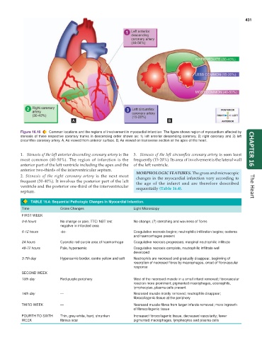
Figure 16.16 Common locations and the regions of involvement in myocardial infarction The figure shows region of myocardium affected by
stenosis of three respective coronary trunks in descending order shown as: 1) left anterior descending coronary, 2) right coronary and 3) left
circumflex coronary artery. A, As viewed from anterior surface. B, As viewed on transverse section at the apex of the heart.
1. Stenosis of the left anterior descending coronary artery is the 3. Stenosis of the left circumflex coronary artery is seen least CHAPTER 16
most common (40-50%). The region of infarction is the frequently (15-20%). Its area of involvement is the lateral wall
anterior part of the left ventricle including the apex and the of the left ventricle.
anterior two-thirds of the interventricular septum. MORPHOLOGIC FEATURES. The gross and microscopic
2. Stenosis of the right coronary artery is the next most changes in the myocardial infarction vary according to
frequent (30-40%). It involves the posterior part of the left the age of the infarct and are therefore described
ventricle and the posterior one-third of the interventricular sequentially (Table 16.4). The Heart
septum.
TABLE 16.4: Sequential Pathologic Changes in Myocardial Infarction.
Time Gross Changes Light Microscopy
FIRST WEEK
0-6 hours No change or pale; TTC/ NBT test No change; (?) stretching and waviness of fibres
negative in infarcted area
6-12 hours -do- Coagulative necrosis begins; neutrophilic infiltration begins; oedema
and haemorrhages present
24 hours Cyanotic red-purple area of haemorrhage Coagulative necrosis progresses; marginal neutrophilic infiltrate
48-72 hours Pale, hyperaemic Coagulative necrosis complete, neutrophilic infiltrate well
developed
3-7th day Hyperaemic border, centre yellow and soft Neutrophils are necrosed and gradually disappear, beginning of
resorption of necrosed fibres by macrophages, onset of fibrovascular
response
SECOND WEEK
10th day Red-purple periphery Most of the necrosed muscle in a small infarct removed; fibrovascular
reaction more prominent; pigmented macrophages, eosinophils,
lymphocytes, plasma cells present
14th day — Necrosed muscle mostly removed; neutrophils disappear;
fibrocollagenic tissue at the periphery
THIRD WEEK — Necrosed muscle fibres from larger infarcts removed; more ingrowth
of fibrocollagenic tissue
FOURTH TO SIXTH Thin, grey-white, hard, shrunken Increased fibrocollagenic tissue, decreased vascularity; fewer
WEEK fibrous scar pigmented macrophages, lymphocytes and plasma cells

