


Page 446 - Textbook of Pathology, 6th Edition
P. 446
430 1. Myocardial ischaemia. Myocardial ischaemia is brought
about by one or more of the following mechanisms:
i) Diminised coronary blood flow e.g. in coronary artery
disease, shock.
ii) Increased myocardial demand e.g. in exercise, emotions.
iii) Hypertrophy of the heart without simultaneous increase
of coronary blood flow e.g. in hypertension, valvular heart
disease.
2. Role of platelets. Rupture of an atherosclerotic plaque
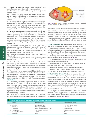
exposes the subendothelial collagen to platelets which Figure 16.15 Diagrammatic representation of extent of myocardial
undergo aggregation, activation and release reaction. These infarction in the depth of myocardium.
events contribute to the build-up of the platelet mass that
may give rise to emboli or initiate thrombosis. but without critical stenosis (not necessarily 75% compro-
3. Acute plaque rupture. In general, slowly-developing mised lumen), aortic stenosis or haemorrhagic shock. This is
coronary ischaemia from stenosing coronary atherosclerosis because subendocardial myocardium is normally least well
of high-grade may not cause acute MI but continue to perfused by coronaries and thus is more vulnerable to any
produce episodes of angina pectoris. But acute complications reduction in the coronary flow. Superimposed coronary
in coronary atherosclerotic plaques in the form of thrombosis is frequently encountered in these cases too, and
superimposed coronary thrombosis due to plaque rupture hence the beneficial role of fibrinolytic treatment in such
and plaque haemorrhage is frequently encountered in cases patients.
of acute MI:
i) Superimposed coronary thrombosis due to disruption of TYPES OF INFARCTS. Infarcts have been classified in a
plaque is seen in about half the cases of acute MI. Infusion of number of ways by the physicians and the pathologists:
intracoronary fibrinolysins in the first half an hour of 1. According to the anatomic region of the left ventricle invol-
development of acute MI in such cases restores blood flow ved, they are called anterior, posterior (inferior), lateral, septal
in the blocked vessel in majority of cases. and circumferential, and their combinations like antero-
ii) Intramural haemorrhage is found in about one-third cases lateral, posterolateral (or inferolateral) and anteroseptal.
of acute MI. 2. According to the degree of thickness of the ventricular wall
Plaque haemorrhage and thrombosis may occur together involved, infarcts are of two types (Fig. 16.15):
in some cases. i) Full-thickness or transmural, when they involve the entire
SECTION III
4. Non-atherosclerotic causes. About 10% cases of acute MI thickness of the ventricular wall.
are caused by non-atherosclerotic factors such as coronary ii) Subendocardial or laminar, when they occupy the inner
vasospasm, arteritis, coronary ostial stenosis, embolism, subendocardial half of the myocardium.
thrombotic diseases, trauma and outside compression as 3. According to the age of infarcts, they are of two types:
already described. i) Newly-formed infarcts called as acute, recent or fresh.
5. Transmural versus subendocardial infarcts. There are ii) Advanced infarcts called as old, healed or organised.
some differences in the pathogenesis of the transmural infarcts
involving the full thickness of ventricular wall and the LOCATION OF INFARCTS. Infarcts are most frequently
subendocardial (laminar) infarcts affecting the inner located in the left ventricle. Right ventricle is less susceptible
subendocardial one-third to half. These are as under to infarction due to its thin wall, having less metabolic
Systemic Pathology
(Table 16.3): requirements and is thus adequately nourished by the
i) Transmural (full thickness) infarcts are the most common thebesian vessels. Atrial infarcts, whenever present, are more
type seen in 95% cases. Critical coronary narrowing (more often in the right atrium, usually accompanying the infarct
than 75% compromised lumen) is of great significance in the of the left ventricle. Left atrium is relatively protected from
causation of such infarcts. Atherosclerotic plaques with infarction because it is supplied by the oxygenated blood in
superimposed thrombosis and intramural haemorrhage are the left atrial chamber.
significant in about 90% cases, and non-atherosclerotic causes The region of infarction depends upon the area of
in the remaining 10% cases. obstructed blood supply by one or more of the three coro-
ii) Subendocardial (laminar) infarcts have their genesis in nary arterial trunks. Accordingly, there are three regions of
reduced coronary perfusion due to coronary atherosclerosis myocardial infarction (Fig. 16.16):
TABLE 16.3: Contrasting Features of Subendocardial and Transmural Infarcts.
Feature Transmural Infarct Subendocardial Infarct
1. Definition Full-thickness, solid Inner third to half, patchy
2. Frequency Most frequent (95%) Less frequent
3. Distribution Specific area of coronary supply Circumferential
4. Pathogenesis > 75% coronary stenosis Hypoperfusion of myocardium
5. Coronary thrombosis Common Rare
6. Epicarditis Common None

