


Page 448 - Textbook of Pathology, 6th Edition
P. 448
432   Grossly, most infarcts occur singly and vary in size from
            4 to 10 cm. As explained above, they are found most often
            in the left ventricle. Less often, there are multifocal lesions.
            The transmural infarcts, which by definition involve the
            entire thickness of the ventricular wall, usually have a thin
            rim of preserved subendocardial myocardium which is
            perfused directly by the blood in the ventricular chamber.
            The subendocardial infarcts which affect the inner sub-
            endocardial half of the myocardium produce less well-
            defined gross changes than the transmural infarcts. The
            sequence of macroscopic changes in all myocardial infarcts
            is as under:
            1. In 6 to 12 hours old infarcts, no striking gross changes
            are discernible except that the affected myocardium is
            slightly paler and drier than normal. However, the early
            infarcts (3 to 6 hours old) can be detected by histochemical
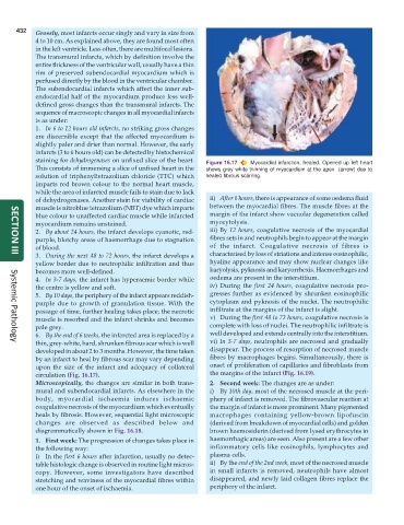
            staining for dehydrogenases on unfixed slice of the heart.  Figure 16.17  Myocardial infarction, healed. Opened up left heart
            This consists of immersing a slice of unfixed heart in the  shows grey white thinning of myocardium at the apex  (arrow) due to
            solution of triphenyltetrazolium chloride (TTC) which  healed fibrous scarring.
            imparts red brown colour to the normal heart muscle,
            while the area of infarcted muscle fails to stain due to lack
            of dehydrogenases. Another stain for viability of cardiac  ii) After 6 hours, there is appearance of some oedema fluid
            muscle is nitroblue tetrazolium (NBT) dye which imparts  between the myocardial fibres. The muscle fibres at the
            blue colour to unaffected cardiac muscle while infarcted  margin of the infarct show vacuolar degeneration called
            myocardium remains unstained.                        myocytolysis.
            2. By about 24 hours, the infarct develops cyanotic, red-  iii) By 12 hours, coagulative necrosis of the myocardial
            purple, blotchy areas of haemorrhage due to stagnation  fibres sets in and neutrophils begin to appear at the margin
            of blood.                                            of the infarct. Coagulative necrosis of fibres is
            3. During the next 48 to 72 hours, the infarct develops a  characterised by loss of striations and intense eosinophilic,
     SECTION III
            yellow border due to neutrophilic infiltration and thus  hyaline appearance and may show nuclear changes like
            becomes more well-defined.                           karyolysis, pyknosis and karyorrhexis. Haemorrhages and
            4. In 3-7 days, the infarct has hyperaemic border while  oedema are present in the interstitium.
            the centre is yellow and soft.                       iv) During the first 24 hours, coagulative necrosis pro-
            5. By 10 days, the periphery of the infarct appears reddish-  gresses further as evidenced by shrunken eosinophilic
            purple due to growth of granulation tissue. With the  cytoplasm and pyknosis of the nuclei. The neutrophilic
            passage of time, further healing takes place; the necrotic  infiltrate at the margins of the infarct is slight.
            muscle is resorbed and the infarct shrinks and becomes  v) During the first 48 to 72 hours, coagulative necrosis is
            pale grey.                                           complete with loss of nuclei. The neutrophilic infiltrate is
            6. By the end of 6 weeks, the infarcted area is replaced by a  well developed and extends centrally into the interstitium.
            thin, grey-white, hard, shrunken fibrous scar which is well  vi) In 3-7 days, neutrophils are necrosed and gradually
     Systemic Pathology
            developed in about 2 to 3 months. However, the time taken  disappear. The process of resorption of necrosed muscle
            by an infarct to heal by fibrous scar may vary depending  fibres by macrophages begins. Simultaneously, there is
            upon the size of the infarct and adequacy of collateral  onset of proliferation of capillaries and fibroblasts from
            circulation (Fig. 16.17).                            the margins of the infarct (Fig. 16.19).
            Microscopically, the changes are similar in both trans-  2. Second week: The changes are as under:
            mural and subendocardial infarcts. As elsewhere in the  i) By 10th day, most of the necrosed muscle at the peri-
            body, myocardial ischaemia induces ischaemic         phery of infarct is removed. The fibrovascular reaction at
            coagulative necrosis of the myocardium which eventually  the margin of infarct is more prominent. Many pigmented
            heals by fibrosis. However, sequential light microscopic  macrophages containing yellow-brown lipofuscin
            changes are observed as described below and          (derived from breakdown of myocardial cells) and golden
            diagrammatically shown in Fig. 16.18.                brown haemosiderin (derived from lysed erythrocytes in
            1. First week: The progression of changes takes place in  haemorrhagic areas) are seen. Also present are a few other
            the following way:                                   inflammatory cells like eosinophils, lymphocytes and
            i) In the first 6 hours after infarction, usually no detec-  plasma cells.
            table histologic change is observed in routine light micros-  ii) By the end of the 2nd week, most of the necrosed muscle
            copy. However, some investigators have described     in small infarcts is removed, neutrophils have almost
            stretching and waviness of the myocardial fibres within  disappeared, and newly laid collagen fibres replace the
            one hour of the onset of ischaemia.                  periphery of the infarct.

