


Page 449 - Textbook of Pathology, 6th Edition
P. 449
433


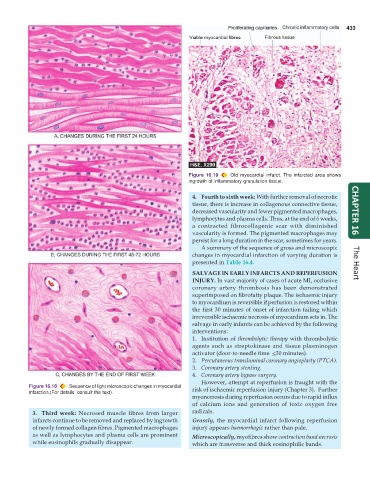
                                                               Figure 16.19  Old myocardial infarct. The infarcted area shows
                                                               ingrowth of inflammatory granulation tissue.

                                                                 4. Fourth to sixth week: With further removal of necrotic
                                                                 tissue, there is increase in collagenous connective tissue,
                                                                 decreased vascularity and fewer pigmented macrophages,  CHAPTER 16
                                                                 lymphocytes and plasma cells. Thus, at the end of 6 weeks,
                                                                 a contracted fibrocollagenic scar with diminished
                                                                 vascularity is formed. The pigmented macrophages may
                                                                 persist for a long duration in the scar, sometimes for years.
                                                                    A summary of the sequence of gross and microscopic
                                                                 changes in myocardial infarction of varying duration is
                                                                 presented in Table 16.4.                             The Heart

                                                                 SALVAGE IN EARLY INFARCTS AND REPERFUSION
                                                                 INJURY. In vast majority of cases of acute MI, occlusive
                                                                 coronary artery thrombosis has been demonstrated
                                                                 superimposed on fibrofatty plaque. The ischaemic injury
                                                                 to myocardium is reversible if perfusion is restored within
                                                                 the first 30 minutes of onset of infarction failing which
                                                                 irreversible ischaemic necrosis of myocardium sets in. The
                                                                 salvage in early infarcts can be achieved by the following
                                                                 interventions:
                                                                 1. Institution of thrombolytic therapy with thrombolytic
                                                                 agents such as streptokinase and tissue plasminogen
                                                                 activator (door-to-needle time  <30 minutes).
                                                                 2. Percutaneous transluminal coronary angioplasty (PTCA).
                                                                 3. Coronary artery stenting.
                                                                 4. Coronary artery bypass surgery.
                                                                    However, attempt at reperfusion is fraught with the
           Figure 16.18  Sequence of light microscopic changes in myocardial  risk of ischaemic reperfusion injury (Chapter 3).  Further
           infarction.(For details, consult the text).
                                                                 myonecrosis during reperfusion occurs due to rapid influx
                                                                 of calcium ions and generation of toxic oxygen free
            3. Third week: Necrosed muscle fibres from larger    radicals.
            infarcts continue to be removed and replaced by ingrowth  Grossly,  the myocardial infarct following reperfusion
            of newly formed collagen fibres. Pigmented macrophages  injury appears haemorrhagic rather than pale.
            as well as lymphocytes and plasma cells are prominent  Microscopically, myofibres show contraction band necrosis
            while eosinophils gradually disappear.               which are transverse and thick eosinophilic bands.

