


Page 494 - Textbook of Pathology, 6th Edition
P. 494
478 iv) It causes considerable obstruction of small airways.
v) It stimulates the vagus and causes bronchoconstriction.
2. Atmospheric pollution. The incidence of chronic
bronchitis is higher in industrialised urban areas where air
is polluted. Some of the atmospheric pollutants which
increase the risk of developing chronic bronchitis are sulfur
dioxide, nitrogen dioxide, particulate dust and toxic fumes.
3. Occupation. Workers engaged in certain occupations
such as in cotton mills (byssinosis), plastic factories etc. are
exposed to various organic or inorganic dusts which
contribute to disabling chronic bronchitis in such individuals.
4. Infection. Bacterial, viral and mycoplasmal infections do
not initiate chronic bronchitis but usually occur secondary
to bronchitis. Cigarette smoke, however, predisposes to
infection responsible for acute exacerbation in chronic
bronchitis.
5. Familial and genetic factors. There appears to be a
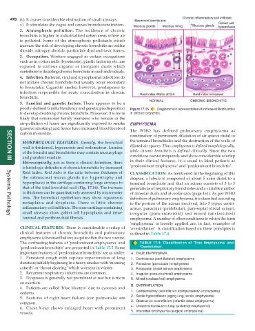
poorly-defined familial tendency and genetic predisposition Figure 17.16 Diagrammatic representation of increased Reid’s index
to develop disabling chronic bronchitis. However, it is more in chronic bronchitis.
likely that nonsmoker family members who remain in the
air-pollution of home are significantly exposed to smoke EMPHYSEMA
(passive smoking) and hence have increased blood levels of The WHO has defined pulmonary emphysema as
carbon monoxide.
combination of permanent dilatation of air spaces distal to
MORPHOLOGIC FEATURES. Grossly, the bronchial the terminal bronchioles and the destruction of the walls of
wall is thickened, hyperaemic and oedematous. Lumina dilated air spaces. Thus, emphysema is defined morphologically,
of the bronchi and bronchioles may contain mucus plugs while chronic bronchitis is defined clinically. Since the two
and purulent exudate. conditions coexist frequently and show considerable overlap
Microscopically, just as there is clinical definition, there in their clinical features, it is usual to label patients as
SECTION III
is histologic definition of chronic bronchitis by increased ‘predominant emphysema’ and ‘predominant bronchitis’.
Reid index. Reid index is the ratio between thickness of CLASSIFICATION. As mentioned in the beginning of this
the submucosal mucus glands (i.e. hypertrophy and chapter, a lobule is composed of about 5 acini distal to a
hyperplasia) in the cartilage-containing large airways to terminal bronchiole and that an acinus consists of 3 to 5
that of the total bronchial wall (Fig. 17.16). The increase generations of respiratory bronchioles and a variable number
in thickness can be quantitatively assessed by micrometer of alveolar ducts and alveolar sacs (page 461). As per WHO
lens. The bronchial epithelium may show squamous definition of pulmonary emphysema, it is classified according
metaplasia and dysplasia. There is little chronic to the portion of the acinus involved, into 5 types: centri-
inflammatory cell infiltrate. The non-cartilage containing acinar, panacinar (panlobular), para-septal (distal acinar),
small airways show goblet cell hyperplasia and intra- irregular (para-cicatricial) and mixed (unclassified)
luminal and peribronchial fibrosis. emphysema. A number of other conditions to which the term
Systemic Pathology
‘emphysema’ is loosely applied are, in fact, examples of
CLINICAL FEATURES. There is considerable overlap of ‘overinflation’. A classification based on these principles is
clinical features of chronic bronchitis and pulmonary outlined in Table 17.4.
emphysema (discussed below) as quite often the two coexist.
The contrasting features of ‘predominant emphysema’ and TABLE 17.4: Classification of ‘True Emphysema’ and
‘predominant bronchitis’ are presented in Table 17.5. Some ‘Overinflation’.
important features of ‘predominant bronchitis’ are as under: A. TRUE EMPHYSEMA
1. Persistent cough with copious expectoration of long 1. Centriacinar (centrilobular) emphysema
duration; initially beginning in a heavy smoker with ‘morning 2. Panacinar (panlobular) emphysema
catarrh’ or ‘throat clearing’ which worsens in winter. 3. Paraseptal (distal acinar) emphysema
2. Recurrent respiratory infections are common. 4. Irregular (para-cicatricial) emphysema
3. Dyspnoea is generally not prominent at rest but is more 5. Mixed (unclassified) emphysema
on exertion.
4. Patients are called ‘blue bloaters’ due to cyanosis and B. OVERINFLATION
oedema. 1. Compensatory overinflation (compensatory emphysema)
5. Features of right heart failure (cor pulmonale) are 2. Senile hyperinflation (aging lung, senile emphysema)
common. 3. Obstructive overinflation (infantile lobar emphysema)
6. Chest X-ray shows enlarged heart with prominent 4. Unilateral translucent lung (unilateral emphysema)
vessels. 5. Interstitial emphysema (surgical emphysema)

