


Page 57 - Textbook of Pathology, 6th Edition
P. 57
41  CHAPTER 3


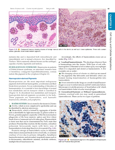
           Figure 3.19  Compound naevus showing clusters of benign naevus cells in the dermis as well as in lower epidermis. These cells contain
           coarse, granular, brown-black melanin pigment.                                                             Cell Injury and Cellular Adaptations


           melanin-like and is deposited both intracellularly and  Accordingly, the effects of haemosiderin excess are as
           intercellularly and is termed ochronosis, first described by  under (Fig. 3.21 ):
           Virchow. Most commonly affected tissues are the cartilages,  a) Localised haemosiderosis.  This develops whenever there
           capsules of joints, ligaments and tendons.          is haemorrhage into the tissues. With lysis of red cells,

           DUBIN-JOHNSON SYNDROME. Hepatocytes in patients     haemoglobin is liberated which is taken up by macrophages
           of Dubin-Johnson syndrome, an autosomal recessive form  where it is degraded and stored as haemosiderin. A few
           of hereditary conjugated hyperbilirubinaemia, contain  examples are as under :
           melain-like pigment in the cytoplasm (Chapter 21).     The changing colours of a bruise or a black eye are caused
                                                               by the pigments like biliverdin and bilirubin which are
           Haemoprotein-derived Pigments                       formed during transformation of haemoglobin into
                                                               haemosiderin.
           Haemoproteins are the most important endogenous        Brown induration in the lungs as a result of small haemor-
           pigments derived from haemoglobin, cytochromes and their  rhages as occur in mitral stenosis and left ventricular failure.
           break-down products. For an understanding of disorders of  Microscopy reveals the presence of ‘heart failure cells’ which
           haemoproteins, it is essential to have knowledge of normal  are haemosiderin-laden alveolar macrophages.
           iron metabolism and its transport which is described in
           Chapter 12. In disordered iron metabolism and transport,  b) Generalised (Systemic or Diffuse) haemosiderosis.
           haemoprotein-derived pigments accumulate in the body.  Systemic overload with iron may result in generalised
           These pigments are haemosiderin, acid haematin      haemosiderosis. There can be two types of patterns:
           (haemozoin), bilirubin, and porphyrins.

           1. HAEMOSIDERIN. Iron is stored in the tissues in 2 forms:
              Ferritin, which is iron complexed to apoferritin and can
           be identified by electron microscopy.
              Haemosiderin, which is formed by aggregates of ferritin
           and is identifiable by light microscopy as golden-yellow to
           brown, granular pigment, especially within the mononuclear
           phagocytes of the bone marrow, spleen and liver where
           break-down of senescent red cells takes place. Haemosiderin
           is ferric iron that can be demonstrated by Perl’s stain that
           produces Prussian blue reaction. In this reaction, colourless
           potassium ferrocyanide reacts with ferric ions of
           haemosiderin to form deep blue ferric-ferrocyanide (Fig. 3.20).
              Excessive storage of haemosiderin occurs in situations
           when there is increased break-down of red cells, or systemic
           overload of iron due to primary (idiopathic, hereditary)
           haemochromatosis, and secondary (acquired) causes such as
           in thalassaemia, sideroblastic anaemia, alcoholic cirrhosis,  Figure 3.20  Haemosiderin pigment in the cytoplasm of hepatocytes
           multiple blood transfusions etc.                    seen as Prussian blue granules.

