


Page 58 - Textbook of Pathology, 6th Edition
P. 58
42 in the hepatocytes. Another variety of haematin pigment is
formalin pigment formed in blood-rich tissues which have been
preserved in acidic formalin solution.
3. BILIRUBIN. Bilirubin is the normal non-iron containing
pigment present in the bile. It is derived from porphyrin ring
of the haem moiety of haemoglobin. Normal level of bilirubin
in blood is less than 1 mg/dl. Excess of bilirubin or hyper-
bilirubinaemia causes an important clinical condition called
jaundice. Normal bilirubin metabolism and pathogenesis of
SECTION I
jaundice are described in Chapter 21. Hyperbilirubinaemia
may be unconjugated or conjugated, and jaundice may
appear in one of the following 3 ways:
a) Prehepatic or haemolytic, when there is excessive destruc-
tion of red cells.
b) Posthepatic or obstructive, which results from obstruction
to the outflow of conjugated bilirubin.
c) Hepatocellular that results from failure of hepatocytes to
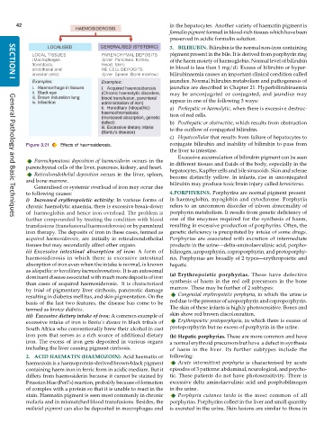
Figure 3.21 Effects of haemosiderosis. conjugate bilirubin and inability of bilirubin to pass from
the liver to intestine.
Excessive accumulation of bilirubin pigment can be seen
Parenchymatous deposition of haemosiderin occurs in the
parenchymal cells of the liver, pancreas, kidney, and heart. in different tissues and fluids of the body, especially in the
hepatocytes, Kupffer cells and bile sinusoids. Skin and sclerae
Reticuloendothelial deposition occurs in the liver, spleen, become distinctly yellow. In infants, rise in unconjugated
and bone marrow. bilirubin may produce toxic brain injury called kernicterus.
Generalised or systemic overload of iron may occur due
to following causes: 4.PORPHYRINS. Porphyrins are normal pigment present
i) Increased erythropoietic activity: In various forms of in haemoglobin, myoglobin and cytochrome. Porphyria
chronic haemolytic anaemia, there is excessive break-down refers to an uncommon disorder of inborn abnormality of
of haemoglobin and hence iron overload. The problem is porphyrin metabolism. It results from genetic deficiency of
further compounded by treating the condition with blood one of the enzymes required for the synthesis of haem,
General Pathology and Basic Techniques
transfusions (transfusional haemosiderosis) or by parenteral resulting in excessive production of porphyrins. Often, the
iron therapy. The deposits of iron in these cases, termed as genetic deficiency is precipitated by intake of some drugs.
acquired haemosiderosis, are initially in reticuloendothelial Porphyrias are associated with excretion of intermediate
tissues but may secondarily affect other organs. products in the urine—delta-aminolaevulinic acid, porpho-
ii) Excessive intestinal absorption of iron: A form of bilinogen, uroporphyrin, coproporphyrin, and protoporphy-
haemosiderosis in which there is excessive intestinal rin. Porphyrias are broadly of 2 types—erythropoietic and
absorption of iron even when the intake is normal, is known hepatic.
as idiopathic or hereditary haemochromatosis. It is an autosomal
dominant disease associated with much more deposits of iron (a) Erythropoietic porphyrias. These have defective
than cases of acquired haemosiderosis. It is characterised synthesis of haem in the red cell precursors in the bone
by triad of pigmentary liver cirrhosis, pancreatic damage marrow. These may be further of 2 subtypes:
resulting in diabetes mellitus, and skin pigmentation. On the Congenital erythropoietic porphyria, in which the urine is
basis of the last two features, the disease has come to be red due to the presence of uroporphyrin and coproporphyrin.
termed as bronze diabetes. The skin of these infants is highly photosensitive. Bones and
iii) Excessive dietary intake of iron: A common example of skin show red brown discolouration.
excessive intake of iron is Bantu’s disease in black tribals of Erythropoietic protoporphyria, in which there is excess of
South Africa who conventionally brew their alcohol in cast protoporphyrin but no excess of porphyrin in the urine.
iron pots that serves as a rich source of additional dietary (b) Hepatic porphyrias. These are more common and have
iron. The excess of iron gets deposited in various organs a normal erythroid precursors but have a defect in synthesis
including the liver causing pigment cirrhosis. of haem in the liver. Its further subtypes include the
2. ACID HAEMATIN (HAEMOZOIN). Acid haematin or following:
haemozoin is a haemoprotein-derived brown-black pigment Acute intermittent porphyria is characterised by acute
containing haem iron in ferric form in acidic medium. But it episodes of 3 patterns: abdominal, neurological, and psycho-
differs from haemosiderin because it cannot be stained by tic. These patients do not have photosensitivity. There is
Prussian blue (Perl’s) reaction, probably because of formation excessive delta aminolaevulinic acid and porphobilinogen
of complex with a protein so that it is unable to react in the in the urine.
stain. Haematin pigment is seen most commonly in chronic Porphyria cutanea tarda is the most common of all
malaria and in mismatched blood transfusions. Besides, the porphyrias. Porphyrins collect in the liver and small quantity
malarial pigment can also be deposited in macrophages and is excreted in the urine. Skin lesions are similar to those in

