


Page 59 - Textbook of Pathology, 6th Edition
P. 59
variegate porphyria. Most of the patients have associated Inhaled Pigments 43
haemosiderosis with cirrhosis which may eventually develop The lungs of most individuals, especially of those living in
into hepatocellular carcinoma. urban areas due to atmospheric pollutants and of smokers,
Mixed (Variegate) porphyrias. It is rare and combines show a large number of inhaled pigmented materials. The
skin photosensitivity with acute abdominal and neurological
manifestations. most commonly inhaled substances are carbon or coal dust;
others are silica or stone dust, iron or iron oxide, asbestos CHAPTER 3
Lipofuscin (Wear and Tear Pigment) and various other organic substances. These substances may
produce occupational lung diseases called pneumoconiosis
Lipofuscin or lipochrome is yellowish-brown intracellular (Chapter 17). The pigment particles after inhalation are taken
lipid pigment (lipo = fat, fuscus = brown). The pigment is up by alveolar macrophages. Some of the pigment-laden
often found in atrophied cells of old age and hence the name macrophages are coughed out via bronchi, while some settle
‘wear and tear pigment’. It is seen in the myocardial fibres, in the interstitial tissue of the lung and in the respiratory
hepatocytes, Leydig cells of the testes and in neurons in senile bronchioles and pass into lymphatics to be deposited in the
dementia. However, the pigment may, at times, accumulate hilar lymph nodes. Anthracosis (i.e. deposition of carbon
rapidly in different cells in wasting diseases unrelated to particles) is seen in almost every adult lung and generally
aging.
provokes no reaction of tissue injury (Fig. 3.23). However,
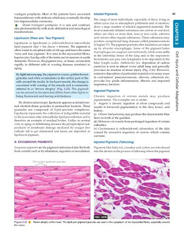
By light microscopy, the pigment is coarse, golden-brown extensive deposition of particulate material over many years
granular and often accumulates in the central part of the in coal-miners’ pneumoconiosis, silicosis, asbestosis etc.
cells around the nuclei. In the heart muscle, the change is provoke low grade inflammation, fibrosis and impaired Cell Injury and Cellular Adaptations
associated with wasting of the muscle and is commonly respiratory function.
referred to as ‘brown atrophy’ (Fig. 3.22). The pigment Ingested Pigments
can be stained by fat stains but differs from other lipids in
being fluorescent and having acid-fastness. Chronic ingestion of certain metals may produce
pigmentation. The examples are as under:
By electron microscopy, lipofuscin appears as intralysoso- i) Argyria is chronic ingestion of silver compounds and
mal electron-dense granules in perinuclear location. These results in brownish pigmentation in the skin, bowel, and
granules are composed of lipid-protein complexes. kidney.
Lipofuscin represents the collection of indigestible material ii) Chronic lead poisoning may produce the characteristic blue
in the lysosomes after intracellular lipid peroxidation and is lines on teeth at the gumline.
therefore an example of residual bodies. Unlike in normal iii) Melanosis coli results from prolonged ingestion of certain
cells, in aging or debilitating diseases the phospholipid end- cathartics.
products of membrane damage mediated by oxygen free iv) Carotenaemia is yellowish-red colouration of the skin
radicals fail to get eliminated and hence are deposited as caused by excessive ingestion of carrots which contain
lipofuscin pigment. carotene.
B. EXOGENOUS PIGMENTS Injected Pigments (Tattooing)
Exogenous pigments are the pigments introduced into the body Pigments like India ink, cinnabar and carbon are introduced
from outside such as by inhalation, ingestion or inoculation. into the dermis in the process of tattooing where the pigment
Figure 3.22 Brown atrophy of the heart. The lipofuscin pigment granules are seen in the cytoplasm of the myocardial fibres, especially around
the nuclei.

