


Page 60 - Textbook of Pathology, 6th Edition
P. 60
44


     SECTION I


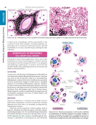
           Figure 3.23  Anthracosis lung. There is presence of abundant coarse black carbon pigment in the septal walls and around the bronchiole.

           is taken up by macrophages and lies permanently in the
           connective tissue. The examples of injected pigments are
           prolonged use of ointments containing mercury, dirt left
           accidentally in a wound, and tattooing by pricking the skin
           with dyes.

                 MORPHOLOGY OF IRREVERSIBLE

                     CELL INJURY (CELL DEATH)
           Cell death is a state of irreversible injury. It may occur in the
           living body as a local or focal change (i.e. autolysis, necrosis
           and apoptosis) and the changes that follow it (i.e. gangrene
     General Pathology and Basic Techniques
           and pathologic calcification), or result in end of the life
           (somatic death). These pathologic processes involved in cell
           death are described below.
           AUTOLYSIS
           Autolysis (i.e. self-digestion) is disintegration of the cell by its
           own hydrolytic enzymes liberated from lysosomes. Autolysis
           can occur in the living body when it is surrounded by
           inflammatory reaction (vital reaction), but the term is generally
           used for postmortem change in which there is complete
           absence of surrounding inflammatory response. Autolysis
           is rapid in some tissues rich in hydrolytic enzymes such as in
           the pancreas, and gastric mucosa; intermediate in tissues like
           the heart, liver and kidney; and  slow in fibrous tissue.
           Morphologically, autolysis is identified by homogeneous and
           eosinophilic cytoplasm with loss of cellular details and
           remains of cell as debris.

           NECROSIS
           Necrosis is defined as a localised area of death of tissue
           followed by degradation of tissue by hydrolytic enzymes
           liberated from dead cells; it is invariably accompanied by
           inflammatory reaction.
              Necrosis can be caused by various agents such as
           hypoxia, chemical and physical agents, microbial agents,
           immunological injury, etc. Two essential changes characterise  Figure 3.24  Necrosis and apoptosis. A, Cell necrosis is identified
           irreversible cell injury in necrosis of all types (Fig. 3.24,A):  by homogeneous, eosinophilic cytoplasm and nuclear changes of
                                                               pyknosis, karyolysis, and karyorrhexis. B, Apoptosis consists of
           i)Cell digestion by lytic enzymes.  Morphologically this  condensation of nuclear chromatin and fragmentation of the cell into
           change is identified as homogeneous and intensely   membrane-bound apoptotic bodies which are engulfed by macrophages.

