


Page 61 - Textbook of Pathology, 6th Edition
P. 61
eosinophilic cytoplasm. Occasionally, it may show   hydrolytic enzymes. The common examples are infarct brain  45
           cytoplasmic vacuolation or dystrophic calcification.  and abscess cavity.
           ii) Denaturation of proteins. This process is morphologically  Grossly, the affected area is soft with liquefied centre
           seen as characteristic nuclear changes in necrotic cell. These  containing necrotic debris. Later, a cyst wall is formed.
           nuclear changes may include: condensation of nuclear
           chromatin (pyknosis) which may either undergo dissolution  Microscopically, the cystic space contains necrotic cell  CHAPTER 3
           (karyolysis) or fragmentation into many granular clumps  debris and macrophages filled with phagocytosed
                                                                 material. The cyst wall is formed by proliferating
           (karyorrhexis) (see Fig. 3.7).
                                                                 capillaries, inflammatory cells, and gliosis (proliferating
                                                                 glial cells) in the case of brain and proliferating fibroblasts
           Types of Necrosis
                                                                 in the case of abscess cavity (Fig. 3.26).
           Morphologically, there are five types of necrosis: coagulative,
           liquefaction (colliquative), caseous, fat, and fibrinoid necrosis.  3. CASEOUS NECROSIS.  Caseous necrosis is found in the
                                                               centre of foci of tuberculous infections. It combines features
           1. COAGULATIVE NECROSIS. This is the most common    of both coagulative and liquefactive necrosis.
           type of necrosis caused by irreversible focal injury, mostly
           from sudden cessation of blood flow (ischaemia), and less  Grossly, foci of caseous necrosis, as the name implies,
           often from bacterial and chemical agents. The organs  resemble dry cheese and are soft, granular and yellowish.
           commonly affected are the heart, kidney, and spleen.  This appearance is partly attributed to the histotoxic
                                                                 effects of lipopolysaccharides present in the capsule of the
            Grossly, foci of coagulative necrosis in the early stage are  tubercle bacilli, Mycobacterium tuberculosis.  Cell Injury and Cellular Adaptations
            pale, firm, and slightly swollen. With progression, they
            become more yellowish, softer, and shrunken.         Microscopically, the necrosed foci are structureless,
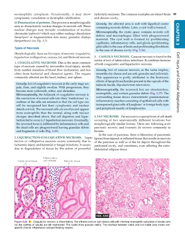
            Microscopically, the hallmark of coagulative necrosis is  eosinophilic, and contain granular debris (Fig. 3.27). The
            the conversion of normal cells into their ‘tombstones’ i.e.  surrounding tissue shows characteristic granulomatous
            outlines of the cells are retained so that the cell type can  inflammatory reaction consisting of epithelioid cells with
            still be recognised but their cytoplasmic and nuclear  interspersed giant cells of Langhans’ or foreign body type
            details are lost. The necrosed cells are swollen and appear  and peripheral mantle of lymphocytes.
            more eosinophilic than the normal, along with nuclear
            changes described above. But cell digestion and lique-  4. FAT NECROSIS.  Fat necrosis is a special form of cell death
            faction fail to occur (c.f. liquefaction necrosis). Eventually,  occurring at two anatomically different locations but
            the necrosed focus is infiltrated by inflammatory cells and  morphologically similar lesions. These are: following acute
            the dead cells are phagocytosed leaving granular debris  pancreatic necrosis, and  traumatic fat necrosis commonly in
            and fragments of cells (Fig. 3.25).                breasts.
                                                                  In the case of pancreas, there is liberation of pancreatic
           2. LIQUEFACTION (COLLIQUATIVE) NECROSIS.  Lique-    lipases from injured or inflamed tissue that results in necrosis
           faction or colliquative necrosis occurs commonly due to  of the pancreas as well as of the fat depots throughout the
           ischaemic injury and bacterial or fungal infections. It occurs  peritoneal cavity, and sometimes, even affecting the extra-
           due to degradation of tissue by the action of powerful  abdominal adipose tissue.


           Figure 3.25  Coagulative necrosis in infarct kidney. The affected area on right shows cells with intensely eosinophilic cytoplasm of tubular cells
           but the outlines of tubules are still maintained. The nuclei show granular debris. The interface between viable and non-viable area shows non-
           specific chronic inflammation and proliferating vessels.

