


Page 62 - Textbook of Pathology, 6th Edition
P. 62
46


     SECTION I


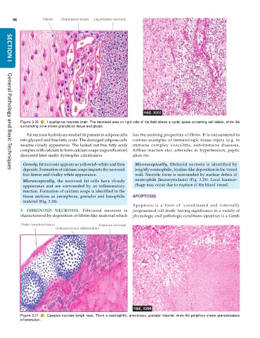
           Figure 3.26  Liquefactive necrosis brain. The necrosed area on right side of the field shows a cystic space containing cell debris, while the
           surrounding zone shows granulation tissue and gliosis.

              Fat necrosis hydrolyses neutral fat present in adipose cells  has the staining properties of fibrin. It is encountered in
           into glycerol and free fatty acids. The damaged adipose cells  various examples of immunologic tissue injury (e.g. in
           assume cloudy appearance. The leaked out free fatty acids  immune complex vasculitis, autoimmune diseases,
           complex with calcium to form calcium soaps (saponification)  Arthus reaction etc), arterioles in hypertension, peptic
           discussed later under dystrophic calcification.     ulcer etc.
            Grossly, fat necrosis appears as yellowish-white and firm  Microscopically, fibrinoid necrosis is identified by
            deposits. Formation of calcium soaps imparts the necrosed  brightly eosinophilic, hyaline-like deposition in the vessel
     General Pathology and Basic Techniques
            foci firmer and chalky white appearance.             wall. Necrotic focus is surrounded by nuclear debris of
            Microscopically, the necrosed fat cells have cloudy  neutrophils (leucocytoclasis) (Fig. 3.29). Local haemor-
            appearance and are surrounded by an inflammatory     rhage may occur due to rupture of the blood vessel.
            reaction. Formation of calcium soaps is identified in the
            tissue sections as amorphous, granular and basophilic  APOPTOSIS
            material (Fig. 3.28).
                                                               Apoptosis is a form of ‘coordinated and internally
           5.  FIBRINOID  NECROSIS. Fibrinoid necrosis is      programmed cell death’ having significance in a variety of
           characterised by deposition of fibrin-like material which  physiologic and pathologic conditions (apoptosis is a Greek


           Figure 3.27  Caseous necrosis lymph node. There is eosinophilic, amorphous, granular material, while the periphery shows granulomatous
           inflammation.

