


Page 63 - Textbook of Pathology, 6th Edition
P. 63
47 CHAPTER 3
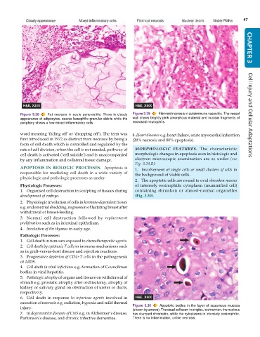
Figure 3.28 Fat necrosis in acute pancreatitis. There is cloudy Figure 3.29 Fibrinoid necrosis in autoimmune vasculitis. The vessel Cell Injury and Cellular Adaptations
appearance of adipocytes, coarse basophilic granular debris while the wall shows brightly pink amorphous material and nuclear fragments of
periphery shows a few mixed inflammatory cells. necrosed neutrophils.
word meaning ‘falling off’ or ‘dropping off’). The term was 8. Heart diseases e.g. heart failure, acute myocardial infarction
first introduced in 1972 as distinct from necrosis by being a (20% necrosis and 80% apoptosis).
form of cell death which is controlled and regulated by the
rate of cell division; when the cell is not needed, pathway of MORPHOLOGIC FEATURES. The characteristic
cell death is activated (‘cell suicide’) and is unaccompanied morphologic changes in apoptosis seen in histologic and
by any inflammation and collateral tissue damage. electron microscopic examination are as under (see
Fig. 3.24,B):
APOPTOSIS IN BIOLOGIC PROCESSES. Apoptosis is 1. Involvement of single cells or small clusters of cells in
responsible for mediating cell death in a wide variety of the background of viable cells.
physiologic and pathologic processes as under:
2. The apoptotic cells are round to oval shrunken masses
Physiologic Processes: of intensely eosinophilic cytoplasm (mummified cell)
1. Organised cell destruction in sculpting of tissues during containing shrunken or almost-normal organelles
development of embryo. (Fig. 3.30).
2. Physiologic involution of cells in hormone-dependent tissues
e.g. endometrial shedding, regression of lactating breast after
withdrawal of breast-feeding.
3. Normal cell destruction followed by replacement
proliferation such as in intestinal epithelium.
4. Involution of the thymus in early age.
Pathologic Processes:
1. Cell death in tumours exposed to chemotherapeutic agents.
2. Cell death by cytotoxic T cells in immune mechanisms such
as in graft-versus-host disease and rejection reactions.
3. Progressive depletion of CD4+T cells in the pathogenesis
of AIDS.
4. Cell death in viral infections e.g. formation of Councilman
bodies in viral hepatitis.
5. Pathologic atrophy of organs and tissues on withdrawal of
stimuli e.g. prostatic atrophy after orchiectomy, atrophy of
kidney or salivary gland on obstruction of ureter or ducts,
respectively.
6. Cell death in response to injurious agents involved in
causation of necrosis e.g. radiation, hypoxia and mild thermal
injury. Figure 3.30 Apoptotic bodies in the layer of squamous mucosa
(shown by arrows). The dead cell seen in singles, is shrunken, the nucleus
7. In degenerative diseases of CNS e.g. in Alzheimer’s disease, has clumped chromatin, while the cytoplasms in intensely eosinophilic.
Parkinson’s disease, and chronic infective dementias. There is no inflammation, unlike necrosis.

