
Page 692 - Textbook of Pathology, 6th Edition
P. 692
676
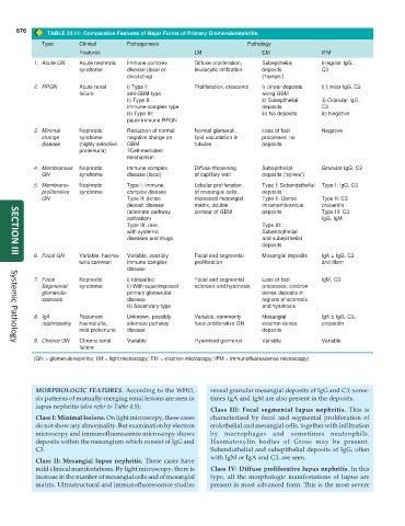
TABLE 22.11: Comparative Features of Major Forms of Primary Glomerulonephritis.
Type Clinical Pathogenesis Pathology
Features LM EM IFM
1. Acute GN Acute nephrotic Immune complex Diffuse proliferation, Subepithelial Irregular IgG,
syndrome disease (local or leucocytic infiltration deposits C3
circulating) (‘humps’)
2. RPGN Acute renal i) Type I: Proliferation, crescents i) Linear deposits i) Linear IgG, C3
failure anti-GBM type along GBM
ii) Type II: ii) Subepithelial ii) Granular IgG,
immune complex type deposits C3
iii) Type III: iii) No deposits iii) Negative
pauci-immune RPGN
3. Minimal Nephrotic Reduction of normal Normal glomeruli, Loss of foot Negative
change syndrome negative charge on lipid vacuolation in processes, no
disease (highly selective GBM tubules deposits
proteinuria) ?Cell-mediated
mechanism
4. Membranous Nephrotic Immune complex Diffuse thickening Subepithelial Granular IgG, C3
GN syndrome disease (local) of capillary wall deposits (‘spikes’)
5. Membrano- Nephrotic Type I: immune Lobular proliferation Type I: Subendothelial Type I: IgG, C3
proliferative syndrome complex disease of mesangial cells, deposits
GN Type II: dense increased mesangial Type II: Dense Type II: C3
deposit disease matrix, double intramembranous properdin
(alternate pathway contour of GBM deposits Type III: C3
activation) IgG, IgM
Type III: rare, Type III:
with systemic Subendothelial
diseases and drugs and subepithelial
deposits
6. Focal GN Variable, haema- Variable, possibly Focal and segmental Mesangial deposits IgA ± IgG, C3
SECTION III
turia common immune complex proliferation and fibrin
disease
7. Focal Nephrotic i) Idiopathic Focal and segmental Loss of foot IgM, C3
Segmental syndrome ii) With superimposed sclerosis and hyalinosis processes, electron
glomerulo- primary glomerular dense deposits in
sclerosis disease regions of sclerosis
iii) Secondary type and hyalinosis
8. IgA Recurrent Unknown, possibly Variable, commonly Mesangial IgA ± IgG, C3,
nephropathy haematuria, alternate pathway focal proliferative GN electron-dense properdin
mild proteinuria disease deposits
9. Chronic GN Chronic renal Variable Hyalinised glomeruli Variable Variable
Systemic Pathology
failure
(GN = glomerulonephritis; LM = light microscopy; EM = electron microscopy; IFM = immunofluorescence microscopy)
MORPHOLOGIC FEATURES. According to the WHO, reveal granular mesangial deposits of IgG and C3; some-
six patterns of mutually-merging renal lesions are seen in times IgA and IgM are also present in the deposits.
lupus nephritis (also refer to Table 4.9):
Class III: Focal segmental lupus nephritis. This is
Class I: Minimal lesions. On light microscopy, these cases characterised by focal and segmental proliferation of
do not show any abnormality. But examination by electron endothelial and mesangial cells, together with infiltration
microscopy and immunofluorescence microscopy shows by macrophages and sometimes neutrophils.
deposits within the mesangium which consist of IgG and Haematoxylin bodies of Gross may be present.
C3. Subendothelial and subepithelial deposits of IgG, often
with IgM or IgA and C3, are seen.
Class II: Mesangial lupus nephritis. These cases have
mild clinical manifestations. By light microscopy, there is Class IV: Diffuse proliferative lupus nephritis. In this
increase in the number of mesangial cells and of mesangial type, all the morphologic manifestations of lupus are
matrix. Ultrastructural and immunofluorescence studies present in most advanced form. This is the most severe