
Page 693 - Textbook of Pathology, 6th Edition
P. 693
and the most common form of lupus nephritis. There is disease is 40 times more common in patients of end-stage 677
diffuse proliferation of endothelial, mesangial, and renal disease in diabetes mellitus than in non-diabetics and
sometimes epithelial cells, involving most or all glomeruli. more diabetics die from cardiovascular complications than
Electron microscopy shows large electron-dense deposits from uraemia.
in the mesangium and in the subendothelial region which MORPHOLOGIC FEATURES. Diabetic nephropathy
on immunofluorescence are positive for IgG; sometimes encompasses 4 types of renal lesions in diabetes mellitus:
also for IgA or IgM, and C3. diabetic glomerulosclerosis, vascular lesions, diabetic
Class V: Membranous lupus nephritis. These lesions pyelonephritis and tubular lesions (Armanni-Ebstein
resemble those of idiopathic membranous GN. These lesions).
consist of diffuse thickening of glomerular capillary wall 1. DIABETIC GLOMERULOSCLEROSIS. Glomerular
on light microscopy and show subendothelial deposits of lesions in diabetes mellitus are particularly common and
immune complexes containing IgG, IgM and C3 on account for majority of abnormal findings referable to the
ultrastructural studies. Mesangial hypercellularity is kidney.
present in some cases.
Pathogenesis of these lesions in diabetes mellitus is
Class VI: Sclerosing lupus nephritis. This is end-stage explained by following sequential changes: hyper-
kidney of SLE, akin to chronic GN. Most glomeruli are glycaemia → glomerular hypertension → renal hyper-
sclerosed and hyalinised and there may be remnants of perfusion → deposition of proteins in the mesangium →
preceding lesions. glomerulosclerosis → renal failure. In addition, cellular
infiltration in renal lesions in diabetic glomerular lesions
Although in a given case, the lesions in lupus nephririts
fit into one of the classes described above, it is not unusual is due to growth factors, particularly transforming growth
to find overlapping and progressive transformation of lupus factor-β. Strict control of blood glucose level and control
lesions during the course of disease. of systemic hypertension in these patients retards
progression to diabetic nephropathy.
Diabetic Nephropathy Glomerulosclerosis in diabetes may take one of the 2
Renal involvement is an important complication of diabetes forms: diffuse or nodular lesions: CHAPTER 22
mellitus. End-stage kidney with renal failure accounts for i) Diffuse glomerulosclerosis. Diffuse glomerular
deaths in more than 10% of all diabetics. Renal complications lesions are the most common. There is involvement of all
are more severe, develop early and more frequently in parts of glomeruli. The pathologic changes consist of
type 1 (earlier called insulin-dependent) diabetes mellitus thickening of the GBM and diffuse increase in mesangial
(30-40% cases) than in type 2 (earlier termed non-insulin- matrix with mild proliferation of mesangial cells. Various
dependent) diabetics (about 20% cases). A variety of clinical exudative lesions such as capsular hyaline drops and fibrin
syndromes are associated with diabetic nephropathy that caps may also be present (Fig. 22.24,A) Capsular drop is an
includes asymptomatic proteinuria, nephrotic syndrome, eosinophilic hyaline thickening of the parietal layer of
progressive renal failure and hypertension. Cardiovascular Bowman’s capsule and bulges into the glomerular space. The Kidney and Lower Urinary Tract
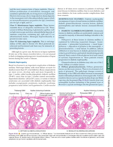
Figure 22.24 Diabetic glomerulosclerosis. A, Diffuse lesions. The characteristic features are dif fuse involvement of the glomeruli showing
thickening of the GBM and diffuse increase in the mesangial matrix with mild proliferation of mesangial cells and exudative lesions (fibrin caps and
capsular drops). B, Nodular lesion (Kimmelstiel-Wilson Lesion). There are one or more hyaline nodules within the lobules of glomeruli, surrounded
peripherally by glomerular capillaries with thickened walls.