
Page 694 - Textbook of Pathology, 6th Edition
P. 694
678 high blood sugar level, the epithelial cells of the proximal
convoluted tubules develop extensive glycogen deposits
appearing as vacuoles. These are called Armanni-Ebstein
lesions. The tubules return to normal on control of
hyperglycaemic state.
Hereditary Nephritis
A group of hereditary diseases principally involving the
glomeruli are termed hereditary nephritis. These include the
following:
1. Alport’s syndrome
2. Fabry’s disease
3. Nail-patella syndrome
1. Alport’s syndrome. Out of various hereditary nephritis,
Alport’s syndrome is relatively more common and has been
extensively studied. This is an X-linked dominant disorder
having mutation in α−5 chain of type IV collagen located on
X-chromosome. It affects males more severely than females.
The syndrome consists of sensori-neural deafness and
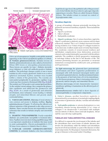
Figure 22.25 Diabetic nephropathy—nodular (Kimmelstiel-W ilson
or KW) lesions. ophthalmic complications (lens dislocation, posterior
cataracts and corneal dystrophy) associated with hereditary
Fibrin cap is homogeneous, brightly eosinophilic material nephritis. The condition is slowly progressive, terminating
appearing on the wall of a peripheral capillary of a lobule. in end-stage kidney in the 2nd to 3rd decades of life. The
ii) Nodular glomerulosclerosis. Nodular lesions of common presenting features are persistent or recurrent
diabetic glomerulosclerosis are also called as Kimmelstiel- haematuria accompanied by erythrocyte casts, proteinuria
Wilson (KW) lesions or intercapillary glomerulosclerosis. and hypertension.
These lesions are specific for type 1 diabetes (juvenile-
onset diabetes) or islet cell antibody-positive diabetes By light microscopy, the glomeruli have predominant
SECTION III
mellitus. The pathologic changes consist of one or more involvement and show segmental proliferation of
nodules in a few or many glomeruli. Nodule is an ovoid or mesangial cells with increased mesangial matrix and
spherical, laminated, hyaline, acellular mass located occasional segmental sclerosis. Another prominent feature
within a lobule of the glomerulus. The nodules are is the presence of lipid-laden foam cells in the interstitium.
surrounded peripherally by glomerular capillary loops As the disease progresses, there is increasing sclerosis of
which may have normal or thickened GBM (Fig. 22.24,B). glomeruli, tubular atrophy and interstitial fibrosis.
The nodules are PAS-positive and contain lipid and fibrin. Electron microscopy reveals characteristic basement
As the nodular lesions enlarge, they compress the glomer- membrane splitting or lamination in the affected parts of
ular capillaries and obliterate the glomerular tuft glomeruli.
(Fig. 22.25). As a result of glomerular and arteriolar Immunofluorescence studies fail to show deposits of
involvement, renal ischaemia occurs leading to tubular immunoglobulins or complement components.
Systemic Pathology
atrophy and interstitial fibrosis and grossly small,
contracted kidney. 2. Fabry’s disease, another hereditary nephritis is
characterised by accumulation of neutral glycosphingolipids
2. VASCULAR LESIONS. Atheroma of renal arteries is in lysosomes of glomerular, tubular, vascular and interstitial
very common and severe in diabetes mellitus. Hyaline cells.
arteriolosclerosis (Chapter 15) affecting the afferent and 3. Nail-patella syndrome or osteonychodysplasia is a rare
efferent arterioles of the glomeruli is also often severe in hereditary disease having abnormality in α-1 chain of
diabetes. These vascular lesions are responsible for renal collagen V on chromosome 9 associated with multiple
ischaemia that results in tubular atrophy and interstitial osseous defects of elbows, knees and nail dysplasia. About
fibrosis.
half the cases develop nephropathy.
3. DIABETIC PYELONEPHRITIS. Poorly-controlled
diabetics are particularly susceptible to bacterial infec- TUBULAR AND TUBULOINTERSTITIAL DISEASES
tions. Papillary necrosis (necrotising papillitis) (page 682)
is an important complication of diabetes that may result It is difficult to separate the involvement of the tubules and
in acute pyelonephritis. Chronic pyelonephritis is 10 to the interstitium since most forms of tubular diseases also
20 times more common in diabetics than in others. involve the interstitium, while the tubules and interstitium
may be involved secondarily as a part of the diseases of other
4. TUBULAR LESIONS (ARMANNI-EBSTEIN renal components. For the purpose of present discussion, this
LESIONS). In untreated diabetics who have extremely
group of diseases is discussed under 2 headings: